Correlating Morphology, Composition, and Optical Behaviour in Group IB and IIB Transition Metal Tartrate Crystals: An SEM–EDAX and UV Study.

Daily writing prompt
Share one of the best gifts you’ve ever received.

               Sachin Nandre1,* Bhushan Nikam2, Hemangi Patil 3

            1,Department of Physics, NSS’S Uttamrao Patil arts and sci.college, Dahiwel Dhule,  India.,

         2 Department of Physics, Kai.Sau.G.F.Patil Jr. College, Shahada Nandurbar, India,

        3,Department of Chemistry, Kai.Sau.G.F.Patil Jr. College, Shahada Nandurbar, India,

 * Author for correspondance (bhushannikam81@gmail.com)

ABSTRACT –

This study presents a systematic correlation between morphology, elemental composition, and optical behaviour of Group IB and IIB transition-metal tartrate crystals investigated using scanning electron microscopy (SEM), energy-dispersive X-ray analysis (EDAX), and ultraviolet (UV) spectroscopy. The combined results establish a strong interdependence between structural characteristics, chemical composition, and optical response, underscoring the significant role of transition metal ions in tailoring the physicochemical properties of tartrate crystals. These findings highlight the potential of such materials for applications in optical and other functional material systems.

Keywords: SEM, EDAX and UV spectroscopic; Optical Behaviour; Transition metal

INTRODUCTION –

Tartrate crystals have attracted significant research interest due to their ferroelectric and piezoelectric properties, as well as their applicability in transducers and both linear and nonlinear mechanical devices. [1-3]. The gel growth technique is one of the simplest and most effective methods for growing sparingly soluble crystals from aqueous solutions, enabling crystal formation under ambient conditions at relatively low temperatures. [4]. Nonlinear optical (NLO) materials play a vital role in optoelectronic applications such as optical frequency conversion, optical data storage, and optical switching in inertial confinement laser fusion systems. To effectively realize these applications, materials exhibiting strong second-order optical nonlinearities, a short transparency cut-off wavelength, and high thermal and mechanical stability are required. [5] In metal–organic coordination complexes, the nonlinear optical (NLO) response is predominantly governed by the organic ligand. With respect to the metallic component, particular attention is given to Group IIB metals (Zn, Cd, Hg, and related ions), as their compounds exhibit high transparency in the ultraviolet region. It is well established that crystal morphology is determined by the interplay between the driving force for crystallization and the diffusion of atoms, ions, molecules, or heat. Variations in these experimental parameters can significantly alter crystal growth behavior, leading to morphological transitions from well-defined polyhedral forms to skeletal and dendrite structures. [6] The growth of single crystals of Calcium tartrate was reported [7] and single crystals of strontium tartrate was reported [8].Thermal studies on tartrate crystals grown by gel method were reported by many investigators [9-11]. Tartrate crystals are of considerable interest, particularly for basic studies of some of their interesting physical properties. Some crystals of this family are ferroelectric [12-14], some others are piezoelectric [15] and quite a few of them have been used for controlling laser emission [16]. The present work investigates the structural, nonlinear, and optical properties of Group IB and IIB transition-metal tartrate crystals, Furthermore, a comprehensive correlation between crystal morphology, elemental composition, and optical behavior was established through combined SEM–EDAX and UV spectroscopic investigations.

MATERIALS AND METHODS –

Raw material for the growth of the tartrate compound was synthesized by mixing aqueous solutions of Tartaric acid (C4H6O6 ) Sodium meta Silicate – Na2SiO3 and  IB and IIB transition metal such as Copper chloride (CuCl2.2H20,), Mercuric chloride (HgCl2) and Cadmium chloride monohydrate (CdCl2.H2O 99 %) with Double distilled water in the amount of specific ratio. The solution was allowed to flow along the test tube wall to prevent cracking of the gel surface. Subsequently, Cu²⁺, Hg²⁺ and Cd²⁺ ions slowly diffused into the gel medium, where they reacted with the inner reactant, resulting in crystal growth.[17] And the corresponding chemical reaction is –

  1. C4H606 + CuCl2→ C4H406Cu + 2HCl
  2. HgCl2 + C4H6O6   →      HgC4H4O6 + 2HCl
  3. C4H6O6 + CdCl2    →     C4H4O6Cd + 2HCl

RESULT AND DISCUSSION

                                                   Fig- SEM , EDAX and UV –Vis

From the optimum growth conditions of copper, mercury, and cadmium tartrate crystals, it is observed that the gel setting time, gel aging time, and crystal growth period vary with different dopants. SEM–EDAX and UV–Vis studies reveal a clear correlation among crystal morphology, elemental composition, and optical behavior.

CHARECTORIZATION STUDY

The crystallographic parameters of the grown crystals were determined from the measured interaxial angles and were found to correspond to orthorhombic and monoclinic crystal systems. UV–Vis spectral analysis revealed that the optical band gap values of all selected Group IB and IIB tartrate crystals are nearly identical, with values of 5.69 eV, 5.87 eV, and 5.85 eV for copper, mercury, and cadmium tartrate crystals, respectively. SEM microstructural analysis indicated distinct surface morphologies for each crystal: copper tartrate exhibited coral reef–like rock structures, mercury tartrate showed coral blossom or octocoral polyp–like features resembling tiny sea flowers, while cadmium tartrate displayed small stone pebble–like and plate–like structures with cut-flower appearances. EDAX analysis confirmed the elemental composition by showing characteristic peaks of copper, mercury, and cadmium, along with silicon, oxygen, carbon, sodium, and chlorine, thereby validating the formation of copper, mercury, and cadmium tartrate crystals.

CONCLUSION

The present study establishes a clear correlation between crystal morphology and the nature of the incorporated transition metal ions in Group IB and IIB tartrate crystals. SEM studies reveal that the crystal morphology of Group IB and IIB metal tartrates is strongly dependent on the type of incorporated metal ion and growth conditions. The observed variations in surface features are well supported by EDAX-confirmed composition and are consistent with the optical behaviour obtained from UV analysis. This correlation highlights the decisive role of metal–ligand interactions in governing the morphological and physicochemical properties of tartrate crystals. Overall, the combined SEM–EDAX–UV analysis demonstrates that crystal morphology is closely linked to elemental composition and plays an important role in governing the optical behavior of transition metal tartrate crystals.

REFRENCES:

[1] R. Mazake ,T. Buslaps ,R.Claessen.J.Fink. Mater. Europhys. Lett.Volume9 (5), pp. 477, 1989

[2] F. Jesu , D. Arivuoli ,S.Ramasamy. Material Resarch Bulletin. Volume 29, Page 309, 1994

[3] M. E. Torres , T. Lopez ,J.Peraza . Journal of Applied Physics. Volume 84, Page 5729, 1998

[4] N. H. Manani , Jethva  Int.Journal of  Scentific research in physics  Lett. Vol. 8, Page 08, 2020

[5] S.Kalaiselvan , G. Pasupathi,B.Sakthivel . Der Pharma Chemica. Volume 4(5), Page 1826, 2012

[6] D. K. Sawant , H.M.Patil ,D.S.Bhavsar,J.H.Patil ,K.D.Girase. Archives of physics Resarch,Volume 2(2) ,Page 67-73, 2011

 [7] S. M. Dharma Prakash , P. Mohan Rao J.. Mater. Sci Lett. Volume 5, Page 769, 1986

 [8] M.H. Rahimkutty, Rajendra Babu ,K. Shreedharan Bull. mater. Sci., Volume 24,Page 249-, 2001.

[9] H.K. Henisch., Crystal growth in gels, University park,PA ; The Pennsylvania university  1973.

[10] P.N. Kotru, N.K. Gupta, K. K. Raina,M.L. Koul, Bull.Mater. Sci. Volume 8 Page 5471986

[11] P.N. Kotru, N.K. Gupta, K. K. RainaL.B.Sarma, Bull. Mater. Sci. Volume 21,Page 83,1986

[12]M. M. Abdel-Kader, FI-Kabbany, S. Taha, M. Abosehly, K. K. Tahoon, and A. EISharkay,

J. Phys. Chem. Sol, Volume 52,Page 655, 1991.

[13] H. B. Gon, J. Cryst. Growth, Volume 102,Page 501,1990

[14] C. C. Desai and A. H. Patel, J. Mat. Sci. Lett, Volume 6, Page 1066, 1987.

[15] V. S. Yadava and V. M. Padmanabhan, Acta. Cryst, B Volume 29,Page 493, 1973.

[16] L. V. Pipree and M. M. Kobklova, Radio Eng. Electron Phys, (USA),Volume 12,Page 33,1984.

[17] B. P. Nikam, S. J. Nandre,C.P.Nikam. JETIR ,Volume 9 (2) , 2022.1984.

Botanical Extracts Used Against Tribolium castaneum (Herbst): A Comprehensive Comparative Review up to 2025

Daily writing prompt
Share one of the best gifts you’ve ever received.

Ravindra N More1, Yuvraj M Bhosale2

1,2PG Department of Zoology, NYNC ACS College, Chalisgaon, Jalgaon 424101 (MH)

Email ID- dryuvrajb0807@gmail.com

ABSTRACT

The red flour beetle, Tribolium castaneum (Herbst), is one of the most destructive cosmopolitan pests of stored grains and processed food products. Its remarkable adaptability, rapid life cycle, and increasing resistance to synthetic fumigants, such as phosphine, have intensified the search for safer and more sustainable alternatives. Botanical extracts, derived from plants rich in bioactive secondary metabolites, have shown promise as environmentally benign methods for controlling pests in stored products. This study offers a thorough, theoretical, and comparative synthesis of plant-derived chemicals, essential oils, and botanical extracts tested against T. castaneum until 2025. The modes of action, effectiveness comparisons, formulation advancements, possibilities for resistance management, and future research goals are highlighted.

To provide a cohesive framework for the logical development of plant-based pesticides for post-harvest protection, this review combines classical and modern literature.

KEYWORDS: Botanical insecticides, Essential oils, Tribolium castaneum, Phytochemicals, Sustainable pest management.

INTRODUCTION

Insects that infest stored products consistently endanger global food security, with Tribolium castaneum being one of the most economically important species because of its capacity to invade flour, cereals, and processed foods (Sokoloff 1974; Campbell and Arbogast 2004). Traditional control methods have relied significantly on chemical fumigants and long-lasting insecticides. Nonetheless, concerns about the environment, food safety problems, and the swift development of resistance, especially to phosphine, have diminished their lasting effectiveness (Coats, 1994; Nayak et al., 2020).

In this context, botanical extracts have received renewed scientific interest. Plants, which have been traditionally employed as grain protectants, possess a wide variety of secondary metabolites that are developed for their defense against herbivores (Fraenkel, 1959; Golob & Webley, 1980; Wink, 2012). Contemporary analytical methods and bioassays have facilitated a thorough assessment of these plants against T. castaneum, uncovering various insecticidal, repellent, antifeedant, and growth-regulating effects (Isman, 2006; Regnault-Roger et al., 2012).

MATERIAL AND METHODS-

  • Biology and Pest Status of Tribolium castaneum

Understanding the biology of T. castaneum is fundamental for evaluating botanical control strategies. The beetle thrives in warm and dry storage conditions and completes multiple generations annually, leading to exponential population growth (Sokoloff, 1974). Both larvae and adult insects can cause quantitative and qualitative losses in food products. They contribute to contamination through the presence of frass (insect droppings), secretions, and allergens (Phillips and Throne, 2010).

Its physiological plasticity and detoxification enzyme systems contribute significantly to insecticide resistance during development (Campbell & Arbogast, 2004; Nayak et al., 2020). These characteristics make T. castaneum an ideal model organism for testing alternative pest control agents, including botanicals with multitarget modes of action.

  • Rationale for Botanical Extracts in Stored-Product Protection

Botanical insecticides offer several advantages over synthetic chemicals, including biodegradability, reduced nontarget toxicity, and a lower risk of resistance development (Isman, 2008; Benelli et al., 2016). Plant-derived compounds often act on multiple physiological pathways, such as neuroreceptors, metabolic enzymes, and hormonal systems, making insect adaptation more difficult (Enan, 2001; Pavela, 2015).

Moreover, many botanicals are locally available and culturally accepted, aligning well with sustainable agriculture and integrated pest management (IPM) frameworks (Dubey et al., 2010; Dubey et al., 2011).

  • Essential Oils as Fumigants and Contact Toxicants

Essential oils represent one of the most extensively studied botanical groups for the control of T. castaneum. Rich in monoterpenoids and phenylpropanoids, these volatile compounds exhibit strong fumigant toxicity, often comparable to synthetic fumigants in laboratory conditions (Lee et al., 2003; Chaubey, 2012).

Mechanistically, essential oils disrupt neural transmission by interacting with octopaminergic receptors and ion channels, leading to paralysis and death (Enan, 2001; Bakkali et al., 2008). Studies have demonstrated high mortality and repellency using oils from Artemisia, Thapsia, and other aromatic plants (Negahban et al., 2007; Salem et al., 2023; Zhang et al., 2024).

  • Plant Powders and Crude Extracts

In addition to essential oils, crude plant powders and solvent extracts have demonstrated significant efficacy against T. castaneum. The leaf and seed powders of Aphanamixis polystachya reduced adult survival and progeny emergence in stored wheat, highlighting the practicality of low-technology applications (Ahmad et al., 2019).

Crude extracts often contain synergistic mixtures of alkaloids, flavonoids, terpenoids, and saponins, which collectively impair feeding, digestion, and reproduction (Harborne, 1998; Wink, 2012). Such complexity may enhance durability against the development of resistance.

  • Saponins and Antinutritional Compounds

The capacity of saponin-rich extracts to damage membranes has drawn attention. Recent studies on Chenopodium quinoa have demonstrated notable insecticidal and antinutritional effects on T. castaneum, linked to midgut injury and digestive enzyme inhibition (El-Sheikh, 2025; Francis et al., 2002).

  • Neem and Classical Botanical Insecticides

Neem (Azadirachta indica) is a benchmark botanical insecticide owing to its broad-spectrum activity and well-characterized mode of action (Schmutterer, 1990). Azadirachtin disrupts molting, reproduction, and feeding behavior in T. castaneum, making it particularly valuable for population suppression rather than rapid knockdown (Isman 2006).

  • Nano Formulations and Technological Advances

Recent advances in nanotechnology have revitalized the research on botanical insecticides. Nanoencapsulation enhances stability, solubility, and controlled release of plant-derived compounds, addressing volatility and degradation issues (Kah et al., 2013).

Although still emerging, nano-formulated botanicals show promises for improving the consistency and scalability of plant-based control strategies against T. castaneum.

  • Comparative Efficacy and Resistance Management

Comparative studies consistently show that while individual botanicals may vary in potency, their multi-site modes of action offer strategic advantages over single-target synthetic insecticides (Pavela & Benelli, 2016; Regnault-Roger et al., 2012).

Importantly, botanicals may play a critical role in resistance management by reducing the selection pressure when integrated with conventional methods (Nayak et al., 2020; Phillips & Throne, 2010).

  • Environmental and Safety Considerations

Botanical insecticides are generally regarded as safer for non-target organisms and consumers, although rigorous toxicological evaluations remain essential (Coats, 1994; Isman, 2020). Their rapid degradation minimizes environmental persistence, which aligns with sustainability goals.

  • Challenges and Future Perspectives

Despite encouraging laboratory findings, challenges such as field validation, standardization, and regulatory acceptance persist (Isman & Grieneisen, 2014; Benelli et al., 2016). Future studies should emphasize formulation science, synergistic mixtures, and practical storage conditions.

CONCLUSION

Botanical extracts are a scientifically valid and eco-friendly option for controlling Tribolium castaneum. Utilizing both conventional wisdom and contemporary studies, these plant-derived solutions provide multifunctional roles, minimize the risk of resistance, and align with sustainable pest control systems. Ongoing interdisciplinary studies are crucial for converting their potential into functional and scalable applications.

Table: Representative botanical extracts evaluated against Tribolium castaneum.

Azadirachta indica (Neem)SeedsAzadirachtin extractGrowth inhibition, reduced fecundityEcdysone disruptionSchmutterer (1990); Isman (2006)
Artemisia sieberi (D. Wormwood)Aerial partsEssential oilHigh fumigant mortalityNeurotoxicityNegahban et al.(2007)
Thapsia garganica (D. Carrots)SeedsEssential oilStrong contact & fumigant toxicityAChE inhibitionSalem et al.(2023)
Chenopodium quinoa (Rajgira)SeedsSaponin-rich extractDigestive inhibitionMembrane disruptionEl-Sheikh (2025)
Aphanamixis polystachya (Pithraj Tree)Leaves & seedsPowderReduced progenyAntifeedantAhmad et al.(2019)

GRAPHICAL ABSTRACT

Plant-derived resources → Extraction (powders, crude extracts, essential oils, nano formulations) → Bioactive phytochemicals (terpenoids, alkaloids, saponins, phenolics) → Multiple physiological targets (nervous system, digestion, reproduction) → Mortality, repellency, population suppression of Tribolium castaneum → Sustainable and residue-safe stored-product protection.

REFERENCES

  1. Abou-Taleb, H. K., El-Sheikh, T. M., & Abdel-Rahman, H. A. (2021). Fumigant toxicity and biochemical effects of selected essential oils   on   the red flour beetle, Tribolium castaneum (Coleoptera: Tenebrionidae).   Journal of Stored   Product   Research, 93, 101825. 
  2. Ahmad, S., Khan, R. R., & Hasan, M. (2019). Insecticidal efficacy of   pithraj (Aphanamixis   polystachya) leaf and seed powders against   Tribolium castaneum   in stored wheat.   Journal of Basic and Applied Zoology, 80(1), 1–10. 
  3. Bakkali, F., Averbeck, S., Averbeck, D., &   Idaomar, M. (2008). Biological effects of essential oils: A review.   Food and Chemical Toxicology, 46(2), 446–475. 
  4. Benelli, G., Pavela, R., Canale, A., Mehlhorn, H., & Murugan, K. (2016). Essential oils as eco-friendly biopesticides Challenges and constraints.   Trends in Plant Science, 21(12), 1000–1007. 
  5. Campbell, J. F., & Arbogast, R. T. (2004). Stored-product insects in changing   climates.   Annual Review of Entomology   49, 351–377. 
  6. Chaubey, M. K. (2012). Biological effects of essential oils   on   stored-product insects.   Journal of Biopesticides, 5(1), 1–10. 
  7. Coats, J. R. (1994). Risks   of   natural versus synthetic insecticides.   Annual Review of Entomology, 39, 489–515. 
  8. Dubey, N. K., Shukla, R., Kumar, A., Singh, P., & Prakash, B. (2010). Global scenario on the application of natural products in integrated pest management   programs.   Journal of Natural Products, 3, 1–18. 
  9. Dubey, N. K., Shukla, R., Kumar, A., Singh, P., & Prakash, B. (2011). Prospects of botanical pesticides in sustainable agriculture.   Current Science, 100(4), 479–488. 
  10. El-Sheikh, T. M. Y. (2025). Antinutritional and insecticidal potential of saponin-rich extract of   Chenopodium quinoa   against   Tribolium castaneum   and its   mechanism of action     Scientific Reports, 15, 10952. 
  11. Enan, E. (2001). Insecticidal activity of essential oils: Octopaminergic sites of action.   Pesticide Biochemistry and Physiology   69(1):   15–22. 
  12. Fraenkel, G. S. (1959). The raison d’être of secondary plant substances.   Science, 129(3361), 1466–1470. 
  13. Francis, G., Kerem, Z., Makkar, H. P. S., & Becker, K. (2002).   Biological     actions   of saponins in animal systems: A review.   British Journal of Nutrition, 88(6), 587–605. 
  14. Golob, P., & Webley, D. J. (1980).   The use of plants and minerals as traditional protectants of stored products   is common. Tropical Products Institute, UK. 
  15. Harborne, J. B. (1998).   Phytochemical methods: A guide to modern techniques of plant analysis (3rd ed.). Springer. 
  16. Isman, M. B. (2006). Botanical insecticides, deterrents and repellents in modern agriculture.   Annual Review of Entomology, 51, 45–66. 
  17. Isman, M. B. (2008). Botanical insecticides: For richer   or   poorer.   Pest   Manag     Sci, 64(1), 8–11. 
  18. Isman, M. B. (2020). Botanical insecticides in the twenty-first century:   Fulfilling their promise?   Annual Review of Entomology, 65, 233–249. 
  19. Isman, M. B., &   Grieneisen, M. L. (2014). Botanical insecticide research: Many publications, limited useful data.   Trends in Plant Science, 19(3), 140–145. 
  20. Kah, M., Beulke, S., Tiede, K., & Hofmann, T. (2013).   Nanopesticides: State of knowledge, environmental fate, and exposure modeling.   Critical Reviews in Environmental Science and Technology, 43(16), 1823–1867. 
  21. Lee, S., Peterson, C. J., & Coats, J. R. (2003). Fumigation toxicity of monoterpenoids to several   stored-product   insects.   Journal of Stored Products Research, 39(1), 77–85. 
  22. Negahban, M.,   Moharramipour, S., &   Sefidkon, F. (2007). Fumigant toxicity of essential oil from   Artemisia   sieberi   against stored-product insects.   Journal of Stored Products Research, 43(2), 123–128. 
  23. Nayak, M. K. Collins, P. J., Pavic, H.,   and   Kopittke, R. A. (2020). Resistance to phosphine in stored-product insects: Current status and future prospects.   Journal of Stored   Product   Research, 86, 101555. 
  24. Papachristos, D. P., &   Stamopoulos, D. C. (2002). Repellent, toxic, and reproduction-inhibitory effects of essential oils on stored-product insects.   Journal of Stored Products Research, 38(2), 117–128. 
  25. Pavela, R. (2015). Acute toxicity and synergistic effects of some monoterpenoid essential oil compounds on   Tribolium castaneum.   Journal of Pest Science, 88(4), 747–754. 
  26. Pavela, R., & Benelli, G. (2016). Essential oils as eco-friendly biopesticides Challenges and constraints:     Industrial Crops and Products, 76, 174–187. 
  27. Phillips, T. W., & Throne, J. E. (2010). Biorational approaches to managing stored-product insects.   Annual Review of Entomology, 55, 375–397. 
  28. Rajendran, S., &   Sriranjini, V. (2008). Plant products as fumigants for stored-product insect control.   Journal of Stored Products Research, 44(2), 126–135. 
  29. Regnault-Roger, C., Vincent, C., & Arnason, J. T. (2012). Essential oils in insect control: Low-risk products in a high-stakes world.   Annual Review of Entomology, 57, 405–424. 
  30. Salem, N.,   Bachrouch, O.,   Sriti, J., & Hammami, M. (2023). Chemical composition and insecticidal activity of   Thapsia     garganica   seed essential oil against   Tribolium castaneum.   Pest Management Science, 79(4), 1562–1571. 
  31. Schmutterer, H. (1990). Properties and potential of natural pesticides from the neem tree.   Annual Review of Entomology, 35, 271–297. 
  32. Sokoloff, A. (1974).   The biology of Tribolium. Oxford University Press. 
  33. Tripathi, A. K., Upadhyay, S., Bhuiyan, M., & Bhattacharya, P. R. (2009). A review on prospects of essential oils as biopesticides.   Current Science, 86(6), 787–794. 
  34. Wink, M. (2012). Plant secondary metabolites as defenses against herbivores.   Annual Plant Reviews, 39, 121–145. 
  35. Zhang, X., Wang, Y., & Liu, Z. (2024). Chemical profiling and insecticidal activity of commercial essential oils against   Tribolium castaneum. Industrial Crops and Products, 210, 118034. 

Evaluation of the Antioxidant properties of Tribulus rajasthanensis L.- A case study

Daily writing prompt
Tell us about your favorite pair of shoes, and where they’ve taken you.

Raju N. Devkar 1 and Dr. Vishal N. Shinde 2

  1. Assistant Professor in Botany, VVM’s S.G. Patil ASC College Sakri Tal. Sakri Dist. Dhule-424304 (MS) India.

Mail ID – rajudevkar094@gmail.com

  • Associate Professor in Botany, ADMSP’s Late Annasaheb R D Deore Art’s and Science College, Mhasadi Tal.Sakri, Dist. Dhule- 424304 (MS) India

Mail ID – vishalshinde1001@gmail.com

ABSTRACT: Free radicals are continuously generated in the body during normal metabolic processes and though exposure to environmental factors such as infectious agents, pollution, UV light and radiations. When these harmful free radicals are not neutralized by primary and secondary defence mechanism of body, oxidative stress occurs, which is the reasons for development of various diseases. Plants have many phytoconstituents including saponin, flavonoids and polyphenol with high antioxidants properties. To determination of antioxidant properties of Tribulus spp. extracts (methanol and aqueous) DPPH (1,1- diphenyl 2- picryl hydrazyl) method was used. Whereas DPPH free radical scavenging activity of methanol extracts revealed the strongest as compared to aqueous extracts.

KEYWORDS: Antioxidants, DPPH, Phenolic compounds, Flavonoids, Tribulus rajasthanensis L.

INTRODUCTION:

          Since ancient times, the medicinal properties of plants have been investigated in the recent scientific developments throughout the world, due to their potent antioxidant activities. As antioxidants have been reported to prevent oxidative damage caused by free radicals, it can interfere with the oxidation process by reacting with free radicals, cheating, catalytic metals and also acting as oxygen scavengers [1]. Reactive oxygen species (ROS), such as hydrogen peroxide (H2O2) and hypochlorous acid (HOCl), and free radicals, such as the superoxide anion (O2) and hydroxyl radical (OH), are produced as normal products of cellular metabolism. Overproduction of free radicals and ROS can lead to oxidative damage to various biomolecules including proteins, lipids, lipoproteins and DNA. This oxidative damage is a critical etiological factor implicated in several chronic disorders such as Cancer, Mellitus, diabetes, inflammatory disease, asthma, cardiovascular disease, neurodegenerative disease and premature aging [2,3]. Antioxidants are means for the substances or group of substances that inhibit oxidative damage to a molecule. This defense system is having many modes of classification such as based on their metabolism of action (chain breaking, preventive). Many plants contain large amounts of antioxidants such as vitamin C, vitamin E, lycopene, lutein, carotenoids, polyphenols which play important roles in adsorbing and neutralizing free radicals [4]. Beside this, phenolic compounds and flavonoids which have been reported to exert multiple biological effects, including free radical scavenging abilities, anti-inflammatory, anticarcinogenic etc. [5].

          Whereas unfavorable environmental conditions for plants, including extreme temperatures, drought, heavy metal exposure, nutrient deficiencies, and high salinity, lead to the excessive production of reactive oxygen species (ROS), which can induce oxidative stress. To counteract this damage, plant cells possess an antioxidant defense system composed of both enzymatic and non-enzymatic components. Non-enzymatic antioxidants act through various mechanisms, such as enzyme inhibition, chelation of trace elements involved in free radical generation, scavenging and neutralization of reactive species, and enhancement of protection via interaction with other antioxidant systems. Among these compounds, secondary metabolites particularly phenolic compounds play a crucial role in protecting plants against oxidative stress [6].

          Tribulus rajasthanensis L. belongs to the family zygophyllaceae. It is an annual plant with a wide global distribution and is commonly found throughout India. The species primarily grows wild in dry and arid regions, especially in West Rajasthan, Gujarat, Maharashtra, Uttar Pradesh, and other similar areas [7, 8, 9].

          The plant is a decumbent herb with pinnately compound leaves. The leaves typically bear 3–10 pairs of sessile leaflets with unequal, oblique, or rounded bases. Flowers are solitary and pentamerous. The number of stamens ranges from five to ten, and the ovary is five-chambered. The fruit is the most characteristic feature of this genus. At maturity, it divides into five indehiscent mericarps, each containing two to five seeds arranged in a horizontal row.

          According to Bhandari and Sharma (1977), the species is closely allied to T. terrestris L. but can be easily distinguished by its secondary spines and the complete absence of lower pair of spines. Typical specimens with mature mericarp can be easily told apart while the intermediate forms that show the characters of both Tribulus rajasthanensis and Tribulus terrestris are difficult to separate. The typical forms of T. rajasthanensis as a variety of T. terrestris [10]. The aim of the present study was to evaluate the antioxidant activity of Tribulus rajasthanensis L. extracts by DPPH methods.

MATERIALS AND METHODS:

Plant materials: The healthy infection free mature plants parts (Fruits, stem, leaves and roots) were collected from the Gomai bank of river, Shahada taluka, Nandurbar District and then they were shade dried and powdered separately in laboratory and kept safely for further research.

Preparation of crude extracts in water: 10 g of dry plant powder was taken in a beaker, 100 ml of distilled water was added, and the mixture was stirred by a magnetic stirrer for 24 h. After that it is filtered by Whatman’s filter paper No.1 and filtrate were centrifuged at 3000 rpm for 15 min. The supernatant was evaporated by rotary evaporator, to get dried form. It was weighed and kept in a refrigerator in sterilized and dark glass containers [11].

Preparation of crude extracts in methanol: Solvent extracts were prepared in methanol at room temperature. 10g of dry plant powder was mixed in sufficient quantity of methanol in conical flask. The conical flasks were plugged tightly with cork. Shaken the conical flask properly to mix the content then kept the conical flask for about 30 minutes for the extraction. After 30 minutes it was filtered and filtrate were collected in china dish. These dishes kept on a water bath for some time to evaporate the solvent, after that the methanolic extract were completely dried.

 Antioxidant Activity (DPPH free radical scavenging activity):

          Free radical scavenging activity was determined using the stable 1,1- diphenyl -2-picryl hydrazyl radical (DPPH) according to the method described by Shimada et al. (1992). Butylated hydroxytoluene (BHT) were used as standard control. Various concentrations of the extracts were added to 4 ml of a 0.004% methanol solution of DPPH. The mixture was shaken and left for 30 minutes at room temperature (25 ± 5⁰C) in the dark, and the absorbance was then measured with a spectrophotometer at 517 nm. All determinations were performed in triplicate [12,13,14]. antioxidant activity was calculated as the percent inhibition caused by the hydrogen donor activity of each sample according to the following:

Inhibition (%) = [(Absorbance control – Absorbance sample)/ (Absorbance control) ×100

Where: absorbance control is the absorbance of DPPH radical plus methanol; absorbance sample is the absorbance of DPPH radical plus sample extract or standard.

RESULTS:

           Many plants exhibits in vitro and in vivo antioxidant properties owing to their phenolics, vitamins, proteins and pectins contents. In the different literatures, it has been revealed that the antioxidant activity of plant extracts is responsible for their therapeutic effect against cancer and many more disorders. Hence, Tribulus rajasthanensis L. plant extracts were evaluated for in vitro antioxidant activities. DPPH (1,1-diphenyl, 2- picryl hydrazyl) method were used for evaluation of in vitro antioxidant activity.

                  In the present study several biochemical constituents and free radical scavenging activity of Tribulus were evaluated. Free radicals are involved in many disorders like neurodegenerative diseases and cancers. Scavenging activity of antioxidants are useful for the control of these diseases. DPPH stable free radical method is a sensitive method to evaluate the antioxidant activity of plant extracts. DPPH radical scavenging activity of methanolic extracts of Tribulus showed strongest while some parts of plants revealed moderate antioxidant properties in aqueous extracts.

DISCUSSION:

           medicinal plants have been used to treat a wide range of disorders since ancient times. From simple cold to complex diseases these plants have served as effective therapeutic agents [15]. Tribulus rajasthanensis L. as a well- known medicinal plant, was selected for this study primarily because of it’s antioxidants potential. Plant extracts were evaluated for in vitro antioxidant activities. DPPH Method provides a good assessment for evaluation of in vitro antioxidant activity. It is based on reaction between antioxidant with nitrogen centered free radical i. e. DPPH (1,1 diphenyl, 2- picryl hydrazyl). That’s why in this experiment; we evaluated the in vitro antioxidant and radical scavenging activities of Tribulus spp. methanol extract using DPPH Method.

           Oxidative stress is a deep-rooted cause of various disorders, including rheumatoid, arthritis and inflammation, neurodegenerative disease, diabetes, cancer, aging etc. Preventing the production of reactive oxygen species (ROS) and reactive nitrogen species (RNS) during cellular metabolism is critically important. The widespread use of medicinal plants across different therapeutic contexts encouraged us to investigate Tribulus spp. to assess its antioxidant and free radical scavenging properties. Our result revealed the tremendous potential of this plant in reducing free radical through DPPH, possibly due to its high polyphenol content. However, more investigations should be carried out to clarify the specific correlations between the plant bioactive and the observed biological activities.

References:

  1. Patel V. R., et al. (2010); Antioxidant activity of some selected medicinal plants in Western region of India. Advances in biological research, 4(1): 23-26.
  2. Ghimire B. K., et al. (2011); A comparative evaluation of the antioxidant activity of some medicinal plants popularly used in Nepal. Journal of medicinal plants research,5(10): 1884-1891.
  3. Patel V. R., et al. (2010); Antioxidant activity of some selected medicinal plants in Western region of India. Advances in biological research, 4(1): 23-26.
  4. Agrawal S. S., et al. (2008); Antioxidant activity of fractions from Tridax procumbens. Journal of Pharmacy research, 2: 71-73.
  5. Patel V. R., et al. (2010); Antioxidant activity of some selected medicinal plants in Western region of India. Advances in biological research, 4(1): 23-26.
  6. Chaves N., et al. (2020); Quantification of the antioxidant activity of plant extracts: Analysis of sensitivity and Hierarchization Based on the method used. MDPI,9(76): 1-15.
  7. Lokhande K. D., et al. (2014); Evaluation of antioxidant potential of Indian wild leafy vegetable Tibullus terrestris. Int J Adv Pharma Biol Chem., 3: 2277- 4688.
  8. Hussain A. A., et al. (2009); study the biological activities of Tribulus terrestris extracts. World Acad Sci Eng Technol., 57: 433-435.
  9. Mohammed M. J. (2008); biological activity of saponins isolated from Tribulus terrestris (fruit) on growth of some bacteria. Tikrit Journal of Pure Science, 13(3): 17-20.
  10. Varghese M., et al. (2006); Taxonomic status of some of the Tribulus species in the Indian subcontinent. Saudi journal of biological sciences, 13(1):7-12.
  11. Abdulqawi L.N. and Syed A.Q. (2021); Evaluation of Antibacterial and Antioxidant activities of Tribulus terrestris L. Fruits. Research J. Pharm. and Tech.,14(1):331-336.
  12. Ghimire B. K., et al. (2011); A comparative evaluation of the antioxidant activity of some medicinal plants popularly used in Nepal. Journal of medicinal plants research,5(10): 1884-1891.
  13. Javed S. R., et al. (2018); In vitro and in Vivo assessment of free radical scavenging and antioxidant activities of Veronica persica Poir. Cellular molecular biology, 57-64.
  14. Patel V. R., et al. (2010); Antioxidant activity of some selected medicinal plants in Western region of India. Advances in biological research, 4(1): 23-26.
  15. Javed S. R., et al. (2018); In vitro and in Vivo assessment of free radical scavenging and antioxidant activities of Veronica persica Poir. Cellular molecular biology, 57-64.

Encapsulation and Polymer Coating Strategies in Autonomous Self-Healing Materials: A Mini Review

Daily writing prompt
Are you patriotic? What does being patriotic mean to you?

Rahul Patil1*, Sunil Sajgane1, Suraj Vasave1, Sandip Patil2

1 Y.C.S.P. Mandal’s Dadasaheb Digambar Shankar Patil Arts, Commerce and Science College, Erandol 425109, Maharashtra, India.    

2 N.T.V.S’s G. T. Patil Arts, Commerce and Science College, Nandurbar 425412, Maharashtra, India.

Corresponding Author Email Id: rahul92ppatil@gmail.com

———————————————————————————————————————

Abstract:

Self-healing materials have garnered significant interest for their ability to autonomously repair damage, improve reliability, and extend the service life of polymer systems. Among various strategies, micro- and nano-encapsulation of healing agents combined with polymeric coatings has emerged as an effective approach, enabling controlled release, protection of active agents, and enhanced mechanical performance. This review highlights recent advances in encapsulation techniques, including physical, chemical, and physico-chemical methods, and examines the influence of capsule size, shell thickness, and morphology on healing efficiency. The selection of polymer coatings thermoset, thermoplastic, and stimuli-responsive is discussed in relation to mechanical reinforcement, environmental resistance, and triggerable release mechanisms. Key self-healing mechanisms, such as capsule rupture, diffusion-based repair, and multi-cycle healing, are summarized. Current challenges, including material compatibility, environmental concerns, cost, and scalability, are addressed, along with future perspectives on sustainable materials, multi-functional coatings, and smart self-healing systems for applications in composites, coatings, electronics, and biomedical devices.

Graphical Abstract:

Keywords: Encapsulation, Self-Healing Coating, Thermoset, Thermoplastic

  1. Introduction:

The growing demand for durable, reliable, and sustainable materials has driven extensive research into self-healing polymer systems capable of autonomously repairing damage and restoring functionality [1,2]. Microcracks generated during service are often precursors to catastrophic failure in polymeric materials and composites, particularly in structural, coating, and electronic applications [3]. Conventional repair strategies are typically labor-intensive, costly, and impractical for inaccessible or microscale damage, motivating the development of materials with intrinsic or extrinsic self-healing capabilities. Among the various self-healing approaches, the encapsulation of healing agents within micro- or nano-sized containers represents one of the most widely investigated and practically viable strategies [2,4]. In encapsulation-based self-healing systems, liquid or solid healing agents are stored within discrete capsules embedded in a polymer matrix. Upon crack initiation and propagation, these capsules rupture or activate, releasing the healing agent into the damaged region where it undergoes polymerization, crosslinking, or physical consolidation, thereby sealing the crack and partially or fully restoring mechanical integrity [5,6].

Micro‑ and nano‑encapsulation offers several advantages over other self‑healing strategies, including effective protection of sensitive healing agents, controlled release behavior, and compatibility with a broad range of polymer matrices [5]. Capsule size plays a critical role in determining healing efficiency, dispersion uniformity, and mechanical performance of the host material. While microcapsules are effective for delivering sufficient quantities of healing agents, nanocapsules provide improved dispersion, reduced stress concentration, and the potential for multiple healing events [7]. Polymer coating or shell materials are a key component of encapsulation‑based self‑healing systems, as they govern capsule stability, mechanical strength, interfacial adhesion, and rupture behavior [2,8]. Commonly employed polymer shells include urea–formaldehyde, melamine–formaldehyde, polyurethane, polyurea, and hybrid shells decorated with inorganic nanolayers for enhanced stability [9]. Recent research has increasingly focused on tailoring polymer coatings through chemical modification or the use of stimuli-responsive polymers to enhance healing efficiency and durability under complex service conditions [8,9]. Despite significant progress, several challenges remain in the large-scale implementation of polymer-coated micro/nano-encapsulation systems, including synthesis scalability, capsule–matrix compatibility, long-term stability, and environmental concerns associated with certain shell materials [2,10]. Therefore, a comprehensive understanding of encapsulation synthesis methods, polymer coating strategies, and their influence on self-healing performance is essential.

This review aims to summarize and critically discuss recent advances in micro- and nano-encapsulation techniques and polymer coating materials used for self-healing applications. Emphasis is placed on synthesis methodologies, structure-property relationships, and practical applications in polymer composites and coatings, while highlighting current limitations and future research directions.

2. Micro/Nano Encapsulation Techniques

Micro- and nano-encapsulation techniques employed for self-healing applications are generally classified into physical, chemical, and physico-chemical methods based on the mechanism of capsule formation. The choice of encapsulation technique significantly influences capsule size, shell morphology, mechanical robustness, and release behavior of the healing agent, thereby affecting overall self-healing efficiency [11,12].

2.1 Physical Methods

Physical encapsulation methods rely primarily on mechanical or thermodynamic processes without involving chemical reactions for shell formation. Common techniques include spray drying, solvent evaporation, phase separation, and melt dispersion [13]. In spray drying, a solution or emulsion containing the healing agent and shell material is atomized into a heated chamber, leading to rapid solvent evaporation and capsule formation. This method is attractive due to its simplicity, scalability, and industrial compatibility; however, it often produces capsules with relatively broad size distributions and limited control over shell thickness [14].

Solvent evaporation and phase separation techniques are widely used for encapsulating liquid healing agents within polymer shells. In these methods, an oil-in-water or water-in-oil emulsion is prepared, followed by controlled solvent removal to induce polymer precipitation around the core material. Although physical methods are cost-effective and easy to implement, the resulting capsules may exhibit lower mechanical strength and reduced stability under long-term service conditions compared to chemically synthesized shells [13].

2.2 Chemical Methods

Chemical encapsulation methods rely on in situ chemical reactions to form polymeric shells around healing agent cores, offering excellent control over capsule size, shell thickness, and mechanical properties, which makes them widely used in self-healing polymer systems [15]. Common approaches include in situ polymerization, interfacial polymerization, and emulsion polymerization. Urea–formaldehyde (UF) and melamine–formaldehyde capsules formed via in situ polymerization exhibit high mechanical strength, thermal stability, and effective rupture during crack propagation [16]. Interfacial polymerization enables the formation of robust polyurethane, polyurea, and polyamide shells, while emulsion polymerization is often employed to produce PMMA shells with uniform morphology. Although chemical methods allow precise tuning of capsule characteristics, the use of toxic monomers and complex reaction conditions raises environmental and safety concerns [15,17].   

Figure 1: Schematic representation of chemical encapsulation methods showing polymeric shell formation around core materials [15,16].

2.3 Physico-Chemical Methods

Physico-chemical encapsulation methods combine elements of both physical and chemical processes to form capsules with tailored properties. Coacervation, sol–gel techniques, and layer-by-layer (LbL) assembly are prominent examples [11,18]. Complex coacervation, based on electrostatic interactions between oppositely charged polymers, enables the formation of capsules with high encapsulation efficiency and relatively uniform size distribution. This method is particularly suitable for temperature-sensitive healing agents.

Sol–gel encapsulation involves the hydrolysis and condensation of inorganic precursors to form hybrid organic–inorganic shells, offering enhanced thermal and chemical stability. Layer-by-layer assembly allows precise control over shell thickness and functionality through sequential deposition of polymeric or inorganic layers, making it attractive for stimuli-responsive and multi-functional self-healing systems. Despite their versatility, physico-chemical methods may face challenges related to processing complexity and scalability [18].

  • Polymer Coating Materials and Strategies

Polymer coatings are widely applied in protective, functional, and controlled-release systems. Selection of coating material and strategy depends on mechanical properties, thermal behavior, and responsiveness to stimuli. Major polymer classes used are thermoset polymers, thermoplastic polymers, and stimuli-responsive polymers.

3.1 Thermoset Polymers

Thermoset polymers are crosslinked materials that form rigid, insoluble, and heat-resistant structures upon curing. The crosslinking process creates a three-dimensional network that imparts excellent mechanical strength, chemical stability, and dimensional integrity. Common examples of thermosets include epoxy resins, polyurethane, and phenolic resins. Due to their robust properties, thermoset polymers are widely employed in protective coatings, corrosion-resistant layers, electrical insulation, adhesives, and even self-healing systems. Their inherent rigidity and resistance to deformation make them ideal for applications where durability under mechanical or chemical stress is critical. However, the irreversible crosslinking reaction also limits their reprocessability; once cured, thermosets cannot be remelted, reshaped, or recycled like thermoplastics. This characteristic necessitates careful processing and design considerations during manufacturing to ensure optimal performance. Advances in thermoset chemistry, including the development of reprocessable or partially reversible networks, are emerging to address these limitations [19].

3.2 Thermoplastic Polymers

In contrast, thermoplastic polymers are linear or slightly branched materials that soften upon heating and solidify when cooled, making them highly processable and recyclable. Common thermoplastics used in coatings include polyethylene (PE), polypropylene (PP), poly(methyl methacrylate) (PMMA), and polyvinyl alcohol (PVA). Their reversible thermal behavior allows for reshaping, extrusion, and molding into complex geometries. Thermoplastic coatings offer advantages such as flexibility, ease of fabrication, and potential for material recovery at the end of life. However, they generally exhibit lower chemical, thermal, and mechanical resistance compared to thermosets, limiting their use in highly demanding environments. Innovations in thermoplastic blends, composites, and nanofiller incorporation aim to improve their performance, particularly for protective and functional coatings [20].  

3.3 Stimuli-Responsive Polymers

Stimuli-responsive or “smart” polymers are materials that undergo reversible changes in their physical or chemical properties in response to external stimuli such as temperature, pH, light, or magnetic fields. For example, poly(N-isopropylacrylamide) (PNIPAM) exhibits thermo-responsive behavior, contracting or swelling with temperature variations, while chitosan and alginate derivatives respond to pH changes for controlled release applications. These polymers are particularly valuable for advanced coating systems, drug delivery platforms, and adaptive surfaces, where dynamic responses to environmental changes are required. By tuning the polymer composition and architecture, it is possible to achieve precise control over release rates, adhesion, permeability, and other functional properties, opening new avenues for smart material design and multifunctional coatings [21].

  • Self-Healing Mechanisms Enabled by Encapsulation

Encapsulation-based self-healing strategies improve the autonomous repair of materials by storing healing agents within micro- or nano-capsules, which are released upon damage. The primary mechanisms include capsule rupture-based healing, diffusion-based healing, and multiple healing cycles.

4.1 Capsule Rupture-Based Healing

Capsule rupture-based self-healing relies on microcapsules embedded within a polymer matrix that release healing agents upon mechanical damage. When a crack propagates through the material, it ruptures the microcapsules, releasing the encapsulated agent into the damaged region. This healing agent subsequently reacts, often in the presence of a catalyst dispersed within the matrix, to polymerize and seal the crack. Commonly used systems include urea-formaldehyde or melamine-formaldehyde microcapsules filled with epoxy, polyurethane, or other reactive monomers. The primary advantage of capsule rupture mechanisms is the rapid, localized repair they provide, which can restore mechanical integrity soon after damage occurs. However, this approach is inherently single-use, as the microcapsules are consumed during the healing event. Once a capsule is depleted, the same site cannot be healed again, limiting the material’s long-term self-healing capability. Researchers have explored methods to improve capsule efficiency and optimize agent loading, ensuring that cracks encounter sufficient healing material to restore strength and prevent crack propagation [22].

4.2 Diffusion-Based Healing

Diffusion-based self-healing strategies rely on the controlled migration of healing agents from internal reservoirs, such as vascular networks, hollow fibers, or nanocapsules, into damaged regions over time. Unlike rupture-based systems, which act only at the moment of mechanical failure, diffusion mechanisms allow continuous, gradual repair and can address more extensive or distributed damage. Healing agents move through the matrix either passively, driven by concentration gradients, or actively in response to external triggers. Integration with stimuli-responsive polymers enhances this approach; for example, changes in temperature, pH, moisture, or light can accelerate or direct the diffusion process. This controlled delivery ensures that healing occurs precisely where and when it is needed, improving durability and extending the operational lifetime of the material [23].

  • Multiple Healing Cycles

Single-use capsule systems are limited by their inability to repair recurrent damage. To address this, multi-capsule arrangements or interconnected microvascular networks have been developed, providing fresh healing agents for repeated healing events. In such systems, cracks can access new reservoirs, enabling multiple repair cycles and significantly prolonging the service life of the polymer. Advanced designs combine capsule and vascular strategies or exploit reversible chemistries, such as Diels–Alder reactions or supramolecular bonding, which allow healing agents to re-form after reaction. These multi-cycle systems are particularly advantageous for high-stress environments or structural applications, where damage may occur repeatedly over time. By integrating both material design and delivery architecture, researchers have created self-healing polymers capable of responding to diverse damage scenarios, making them more practical for industrial and commercial applications [24].

4.4 Comparative Overview of Self-Healing Strategies

Each self-healing approach offers unique advantages and limitations depending on the application. Capsule rupture-based systems provide fast, localized repair and are relatively simple to implement, but their single-use nature restricts long-term effectiveness. Diffusion-based mechanisms, in contrast, allow continuous or delayed healing over larger areas and can be finely tuned using stimuli-responsive polymers; however, their repair rate may be slower, and precise control over agent migration may be challenging [25]. Multi-cycle healing systems address the limitations of single-use capsules by providing repeated access to fresh healing agents, either through microvascular networks or reversible chemistries, enhancing durability and structural longevity. By carefully selecting and combining these strategies, materials can be engineered for specific operational requirements-for instance, rapid localized repair for low-damage risk environments, sustained healing for slow-degrading materials, or multiple-cycle systems for critical load-bearing applications. The ongoing development of hybrid approaches, such as integrating capsule rupture with vascular delivery or embedding smart stimuli-responsive agents, offers the potential to achieve both immediate and long-term self-healing performance, making polymers more reliable and resilient for industrial, aerospace, and biomedical applications [26].

  • Applications in Polymer Composites

Structural Materials:

Diffusion-based self-healing is particularly valuable in structural polymer composites, where the formation of microcracks over time can severely compromise mechanical integrity and lead to premature failure. Unlike single-use capsule systems, diffusion-based mechanisms allow healing agents to gradually migrate into damaged regions, enabling continuous or repeated repair even in areas that are difficult to access. This property is especially important in aerospace, automotive, and civil engineering applications, where components are subject to cyclic loading, environmental degradation, and complex stress distributions. By maintaining the integrity of the polymer matrix, diffusion-based healing reduces the risk of crack coalescence and catastrophic failure, effectively extending the service life of high-performance materials. Advanced designs often integrate stimuli-responsive polymers, where temperature, moisture, or mechanical stress can trigger or accelerate the diffusion of healing agents, ensuring timely repair. Additionally, diffusion-based systems can be combined with fiber-reinforced composites or microvascular networks to optimize the distribution of healing agents throughout large, load-bearing structures, providing both durability and resilience in demanding operational environments [27].

Coatings:

In protective coatings, self-healing polymers play a critical role in restoring barrier properties against environmental degradation, chemical attack, corrosion, or mechanical wear. Diffusion-based mechanisms are particularly advantageous in this context, as they allow healing agents to gradually migrate into damaged or scratched regions without requiring external intervention, maintaining the continuity and integrity of the coating. This ensures that the underlying substrate remains shielded from moisture, oxygen, or corrosive agents, which is especially important in metal structures, pipelines, and marine equipment. Stimuli-responsive coatings further enhance performance by activating healing processes in response to environmental triggers such as changes in moisture, pH, temperature, or even UV exposure. For instance, pH-sensitive coatings can release corrosion inhibitors when exposed to acidic conditions, while moisture-responsive systems can accelerate polymerization to seal microcracks. Incorporating nanocapsules or microvascular networks into these coatings can improve the distribution and availability of healing agents, allowing repeated or localized repairs and prolonging the operational life of protective surfaces in harsh industrial, marine, or infrastructure environments [28].

Electronics & Smart Materials:

Self-healing polymer composites are increasingly being adopted in flexible electronics, wearable devices, sensors, and other smart materials, where mechanical integrity and consistent performance are critical. Diffusion-based healing mechanisms play a key role in these applications by allowing healing agents to migrate into microcracks or damaged regions, restoring both the structural and functional properties of the material without external intervention. This ensures that minor mechanical damages—such as bending, stretching, or accidental scratches do not disrupt electrical pathways or compromise device functionality. When combined with stimuli-responsive polymers, the self-healing process can be precisely triggered by environmental or operational signals, such as temperature changes, light exposure, or electrical currents. Such targeted healing not only repairs physical damage but also preserves conductivity, sensor sensitivity, and overall device performance. Additionally, integrating nanocapsules, conductive fillers, or microvascular networks within the polymer matrix enhances the efficiency and speed of repair, supporting long-term reliability. These diffusion-enabled, smart self-healing systems are particularly important for next-generation electronics, soft robotics, and adaptive materials, where durability, resilience, and uninterrupted functionality are essential under repeated deformation or harsh operating conditions [29].

Figure 2: Applications of Self-Healing Micro/Nano Capsules in Structural Materials, Coatings, Electronics & Smart Materials.  

  • Challenges and Future Perspectives

Despite significant advances in micro/nano encapsulation and self-healing polymer systems, several challenges remain that limit their widespread application. One major issue is the compatibility and stability of the encapsulated agents within the polymer matrix; Premature leakage, aggregation, or chemical degradation of the core material can reduce healing efficiency and long-term performance. Achieving uniform capsule size, shell thickness, and mechanical robustness, particularly at the nanoscale, also remains difficult, directly impacting reproducibility and release kinetics. Additionally, many chemical encapsulation processes rely on toxic monomers or organic solvents, raising environmental and safety concerns. The cost and scalability of producing high-quality micro/nano capsules and self-healing polymers is another limiting factor, preventing large-scale commercialization [30].

Future developments in the field are focused on addressing these limitations. The use of biodegradable polymers, water-based systems, and non-toxic monomers can reduce environmental impact while maintaining functional performance. Integration of stimuli-responsive polymers, multi-agent encapsulation, and nanoengineered shells offers potential for more precise controlled release, repeated healing cycles, and targeted delivery. Advances in fabrication techniques, including microfluidics, 3D printing, and layer-by-layer assembly, promise improved precision, reproducibility, and scalability. Finally, combining self-healing polymers with sensing technologies, electronics, or biomedical devices could enable the development of autonomous, adaptive, and multifunctional materials, paving the way for next-generation applications in a variety of industries.   

  • Conclusion

Micro- and nano-encapsulation of healing agents, combined with tailored polymer coatings, represents a highly effective strategy for developing autonomous self-healing polymer systems. Physical, chemical, and physico-chemical encapsulation techniques allow precise control over capsule size, shell thickness, and release behavior, while thermoset, thermoplastic, and stimuli-responsive coatings enhance mechanical strength, environmental stability, and controlled activation of healing. Capsule rupture, diffusion-driven repair, and multi-cycle healing mechanisms demonstrate the versatility and practical applicability of these systems in polymers, composites, coatings, and biomedical materials. Despite significant progress, challenges such as material compatibility, environmental impact, scalability, and cost remain. Future research should focus on sustainable polymers, multi-functional coatings, and integration with smart and adaptive systems to achieve repeated healing, improved efficiency, and commercial viability. Overall, encapsulation-based self-healing strategies hold great promise for extending the service life and reliability of polymeric materials in diverse applications.

Acknowledgement: Rahul Patil sincerely acknowledges Kavayitri Bahinabai Chaudhari North Maharashtra University, Jalgaon, for awarding the Vice-Chancellor Research Motivation Scheme (VCRMS), which supported the research project.

References:

  1. White, S. R., Sottos, N. R., Geubelle, P. H., Moore, J. S., Kessler, M. R., Sriram, S. R., Viswanathan, S. (2001). Autonomic healing of polymer composites. Nature, 409(6822), 794–797. https://doi.org/10.1038/35057232.
  2. Polymers Special Issue. (2025). Polymer micro‑ and nanocapsules: Current status, challenges, andopportunities.Polymers.https://www.mdpi.com/journal/polymers/special_issues/Polymer_Micro_Nanocapsules.
  3. Nayak, S., Vaidhun, B., & Kedar, K. (2024). Applications of microcapsules in self-healing polymeric materials. Current Nanoscience, 2, 218–241.
  4. Zehra, S., Mobin, M., Aslam, R., & Bhat, S. u. I. (2023). Nanocontainers: A comprehensive review on their application in stimuli-responsive smart functional coatings. Progress in Organic Coatings, 176, 107389. https://doi.org/10.1016/j.porgcoat.2023.107389.
  5. Montemor, M. F. (2024). Advanced micro/nanocapsules for self-healing coatings. Applied Sciences, 14(18), 8396. https://doi.org/10.3390/app14188396.
  6. Yuan, Y., et al. (2023). Self‑healing poly(urea formaldehyde) microcapsules: Synthesis and characterization. Polymers, 15(7), 1668. https://doi.org/10.3390/polym15071668
  7. Jiang, Y., Yao, J., & Zhu, C. (2022). Improving the dispersibility of poly(urea‑formaldehyde) microcapsules for self‑healing coatings using preparation process. Journal of Renewable Materials, 10(1), 135–148. https://doi.org/10.32604/jrm.2021.016304
  8. Polythiourethane microcapsules as novel self‑healing systems for epoxy coatings. (2017). Polymer Bulletin. https://doi.org/10.1007/s00289‑017‑2021‑3.
  9. Montemor, M. F. (2014). Functional and smart coatings for corrosion protection: A review of recent advances. Surface and Coatings Technology, 258, 17–37. https://doi.org/10.1016/j.surfcoat.2014.06.031.
  10. Preparation and properties of melamine urea-formaldehyde microcapsules for self-healing of cementitious materials. (2017). PubMed. https://pubmed.ncbi.nlm.nih.gov/28773280/.
  11. Zhu, Y., Ye, X., Rong, M. Z., & Zhang, M. Q. (2015). Self-healing polymeric materials based on microencapsulated healing agents: From design to preparation. Progress in Polymer Science, 49–50, 175–220. https://doi.org/10.1016/j.progpolymsci.2015.07.002.
  12. Blaiszik, B. J., Kramer, S. L. B., Olugebefola, S. C., Moore, J. S., Sottos, N. R., & White, S. R. (2010). Self-healing polymers and composites. Annual Review of Materials Research, 40, 179–211. https://doi.org/10.1146/annurev-matsci-070909-104532.
  13. Zuidema, J. M., Rivet, C. J., Gilbert, R. J., & Morrison, F. A. (2014). A review of microencapsulation techniques for self-healing materials. Journal of Materials Chemistry A, 2(27), 10964–10977. https://doi.org/10.1039/C4TA00643F.
  14. Gharsallaoui, A., Roudaut, G., Chambin, O., Voilley, A., & Saurel, R. (2007). Applications of spray-drying in microencapsulation of food ingredients: An overview. Food Research International, 40(9), 1107–1121. https://doi.org/10.1016/j.foodres.2007.07.004.
  15. Brown, E. N., White, S. R., & Sottos, N. R. (2004). Microcapsule induced toughening in a self-healing polymer composite. Journal of Materials Science, 39(5), 1703–1710. https://doi.org/10.1023/B:JMSC.0000016173.73733.dc.
  16. Yuan, Y. C., Rong, M. Z., & Zhang, M. Q. (2008). Preparation and characterization of microencapsulated polythiol. Polymer, 49(10), 2531–2541. https://doi.org/10.1016/j.polymer.2008.03.042.
  17. Wu, D. Y., & Meure, S. (2008). Self-healing polymeric materials: A review of recent developments. Progress in Polymer Science, 33(5), 479–522. https://doi.org/10.1016/j.progpolymsci.2008.02.001.
  18. Decher, G. (1997). Fuzzy nanoassemblies: Toward layered polymeric multicomposites. Science, 277(5330), 1232–1237. https://doi.org/10.1126/science.277.5330.1232.
  19. Pascault, J. P., Sautereau, H., Verdu, J., & Williams, R. J. J. (2002). Thermosetting polymers. CRC Press.
  20. Kumar, R., & Varadarajan, K. M. (2015). Thermoplastic polymers: Processing, properties, and applications. Polymer Reviews, 55(3), 395–429. https://doi.org/10.1080/15583724.2015.1029494.
  21. Qiu, Y., & Park, K. (2001). Environment-sensitive hydrogels for drug delivery. Advanced Drug Delivery Reviews, 53(3), 321–339. https://doi.org/10.1016/S0169-409X(01)00203-0.
  22. Brown, E. N., Kessler, M. R., Sottos, N. R., & White, S. R. (2005). In situ poly(urea-formaldehyde) microencapsulation of dicyclopentadiene. Journal of Microencapsulation, 22(6), 619–632. https://doi.org/10.1080/02652040500413581.
  23. Toohey, K. S., Sottos, N. R., Lewis, J. A., Moore, J. S., & White, S. R. (2007). Self-healing materials with microvascular networks. Nature Materials, 6(8), 581–585. https://doi.org/10.1038/nmat1944.
  24. Groves, R. M., Toohey, K. S., White, S. R., & Sottos, N. R. (2009). Vascular-based self-healing polymers. Polymer, 50(5), 1283–1290. https://doi.org/10.1016/j.polymer.2009.01.041.
  25. Liu, B., Wu, M., Du, W., Jiang, L., Li, H., Wang, L., & Ding, Q. (2023). The application of self-healing microcapsule technology in the field of cement-based materials: A review and prospect. Polymers, 15(12), 2718. https://doi.org/10.3390/polym15122718.
  26. Amaral, A. J. R., & Pasparakis, G. (2017). Stimuli responsive self-healing polymers: gels, elastomers and membranes. Polymer Chemistry, 8, 6464–6484. https://doi.org/10.1039/c7py01386h.
  27. 1. Blaiszik, B. J., Kramer, S. L. B., Olugebefola, S. C., Moore, J. S., Sottos, N. R., & White, S. R. (2010). Self healing polymers and composites. Annual Review of Materials Research, 40, 179–211. https://doi.org/10.1146/annurev-matsci-070909-104532.
  28. Zhang, Y., Li, X., & Chen, Z. (2023). Self-healing polymer-based coatings: Mechanisms and applications. Polymers, 17(23), 3154. https://doi.org/10.3390/polym17233154.
  29. Choi, K., Noh, A., Kim, J., Hong, P. H., Ko, M. J., & Hong, S. W. (2023). Properties and applications of self-healing polymeric materials: A review. Polymers, 15(22), 4408. https://doi.org/10.3390/polym15224408.
  30. Stuart, M. A. C., Huck, W. T. S., Genzer, J., Müller, M., Ober, C., Stamm, M., Sukhorukov, G. B., Szleifer, I., Tsukruk, V. V., Urban, M., Winnik, F., Zauscher, S., Luzinov, I., & Minko, S. (2010). Emerging applications of stimuli-responsive polymer materials. Nature Materials, 9(2), 101–113. https://doi.org/10.1038/nmat2614.     

Forest Loss and Sustainable Development Challenges in the Satpuda Fringe of North Maharashtra: A Remote Sensing–Based Assessment

Dr. Sachin Ranu Govardhane

Dept of Geography

V.V. Ms S. G. Patil Arts, Science  And Commerce College Sakri,

Tal- Sakri Dist- Dhule.

Email Id sachingovardhane@gmail.com

Abstract

Forest ecosystems in the Satpuda fringe of North Maharashtra are very crucial in maintaining the ecological stability and tribal livelihoods, but they are becoming under pressure due to development pressures. The paper evaluates the change in forest cover and its sustainability development in the Satpuda fringe in 2015-2025 using a remote sensing and GIS-based methodology. Geometric, radiometric, and atmospheric corrections were applied to multi-temporal satellite images of Landsat 8 (2015) and Landsat 9/Sentinel-2 (2025). In ArcGIS Pro, supervised classification and post-classification change detection methods were used to measure the change of tehsil-wise forest cover in terms of area and percentage. The findings indicate a general and geographically imbalanced decrease in forest cover in the study area. The loss of forest was found to be significant in Akrani (247.90 sq. km; -19.14%) and Akkalkuwa (109.25 sq. km; -11.74%), which means that there is a strong pressure in tribal tehsils with a lot of forest. Other tehsils had moderate declines, with only Raver having a marginal growth (3.42 sq. km; +0.36%), probably because of local afforestation. Forest loss spatial distribution is in close relation to population increase, agricultural activities, and infrastructure. The paper identifies the urgency to have integrated land-use planning and conservation-based development policies to achieve long-term sustainability in the ecologically sensitive Satpuda fringe of North Maharashtra.

Keywords

Forest cover change; Remote sensing and GIS; Change detection; Sustainable development; Land use–land cover; Satpuda fringe, North Maharashtra.

Introduction

Forests are very important in ensuring an ecological balance, rural livelihoods and sustainable development, especially in socio-economically vulnerable and environmentally sensitive areas. In India, forested landscapes situated in hill ranges and tribal belts are becoming more and more strained with the increasing population, agricultural activities, development of infrastructure, and the shift in land-use patterns (Behera et al., 2015). The Satpuda Range, particularly its northern edge into North Maharashtra, including Nandurbar district, Dhule district and Jalgaon district is one such ecologically important area. This area is a transitional area whereby thick forest cover is slowly being replaced by agricultural land and human habitation and is therefore very vulnerable to forest degradation and loss (Zurqani et al., 2019).

The Satpuda fringe is typified by topography that is undulating, a forest cover that is mainly comprised of the deciduous forests and a tribal population that relies heavily on the forest cover to provide fuelwood, fodder, small forest produce and subsistence agriculture (Gautam et al., 2002). In the past 10 years, the traditional land-use patterns have changed due to developmental activities like road construction, agricultural intensification, expansion of settlements, and demographic growth (Giriraj et al., 2008). Although these changes are meant to enhance economic status and infrastructure, they tend to have unplanned ecological effects, especially the loss and degradation of forest cover (Bas et al., 2024). The loss of forests in such areas not only endangers the biodiversity and ecosystem services but also the very basis of sustainable development as it impacts the water availability, soil stability, and livelihood security (Kline et al., 2004).

The economic growth, social well-being, and environmental conservation must be balanced in a careful manner to achieve sustainable development in the forest-dependent regions (Mani & Varghese, 2018). Nevertheless, this balance is difficult to measure without credible and spatially explicit information on forest dynamics and association with development processes (Islomov et al., 2023). In this regard, remote sensing and GIS methods provide an effective and inexpensive method of tracking the change in forest cover over time (Stamatopoulos et al., 2024). The satellite-based analysis can be used to consistently monitor large and inaccessible regions and enable researchers to measure the loss of forests, spatial dynamics, and correlate them with socio-economic factors such as population growth and agricultural development at more specific administrative units like tehsils.

Although national level statistics on forests are available, localized research on current changes and development pressures at tehsil level is scarce in the case of the Satpuda fringe of North Maharashtra (Syamsih, 2024). In response to this gap, the current paper conducts a remote sensing-based evaluation of the change in forest cover between 2015 and 2025, a time when the area is experiencing a high rate of development. The study aims to combine satellite-based forest data with simple development indicators to gain a better insight into the spatial distribution of forest loss and its overlap with the ongoing development processes. The results should be relevant to the regional-level planning by identifying priority areas in which the development strategies should be more aligned with the forest conservation and long-term sustainability objectives.

Study Area

The study site is the Satpuda fringe of North Maharashtra, which is a region in the south foothills of Satpuda Range. It covers portions of Nandurbar district, Dhule district and Jalgaon district, and is a transitional region between forested hills and agricultural plains. The area is also marked by a topography of undulations, tropical dry deciduous forests and a majorly tribal population that relies on forest resources. Over the past years, population growth, agricultural activities, and development of infrastructure have escalated the pressure on forest areas and thus the Satpuda fringe is a vital area to understand the issues of forest loss and sustainable development.

Figure 3 LULC Map of Satpuda fringe of North Maharashtra during 2015–2025 to show change in forest cover.

Aim

The aim of the study is to assess forest loss and its implications for sustainable development in the Satpuda fringe of North Maharashtra by analyzing recent forest cover changes using remote sensing techniques and examining their relationship with selected development indicators.

Objectives

  • To assess changes in forest cover in the Satpuda fringe of North Maharashtra between 2015 and 2025.
  • To identify and analyze the spatial patterns of forest loss at the tehsil level within the study area.
  • To examine the relationship between forest loss and development indicators, particularly population growth and agricultural expansion.
  • To evaluate the implications of forest loss for sustainable development.

Methodology and Database

The current research uses a remote sensing and GIS-based approach to evaluate the change in forest cover and its effects on sustainable development in the Satpuda fringe of North Maharashtra. The decadal changes in the forest cover were analyzed using multi-temporal satellite data of 2015 and 2025 on the tehsil level. Available sources of cloud-free satellite images of NASA included Landsat 8 (OLI) in 2015 and Landsat 9 (OLI-2) or Sentinel-2 in 2025. The images were geometrically fixed, radiometrically fixed, and atmospherically fixed to make them comparable over time. With the assistance of visual interpretation and available forest cover maps, supervised classification methods were used to classify forest and non-forest classes. Post-classification comparison was used to measure change in forest area (sq. km and percent) in the two reference years.

The ArcGIS Pro software was used to perform spatial analysis to compute the tehsil-wise forest cover statistics and to determine the spatial patterns of forest loss. The data on tehsil boundaries were collected through SOI official sources of administration and superimposed on the classified forest maps to derive information on areas. These datasets were combined with spatial outputs in order to understand development-based pressures on forest resources. The integration of satellite imagery, GIS-based spatial analysis, and secondary statistical data will be a strong database to assess forest loss and its impact on sustainable development in the Satpuda fringe of North Maharashtra.

Table 1 Spatial analysis of Forest cover area in the Satpuda fringe of North Maharashtra

Year20152025Change Detection
TehsilArea (Sq.km)Area (%)Area (Sq.km)Area (%)Decrease Area (Sq.km)Increase Area (Sq.km)Decrease Area (%)Increase Area (%)
Akkalkuwa521.47456.04412.22744.30109.24711.74
Akrani1060.54181.90812.64462.75247.89719.14
Taloda99.42621.8791.02020.028.4051.85
Shahada111.6209.45103.3468.758.2740.70
Shirpur363.55624.11337.16722.3626.3891.75
Chopda361.09231.36356.75530.994.3370.38
Yaval306.46033.11288.43031.1618.0301.95
Raver317.36133.78320.78434.143.4240.36

(Source: Calculated by researcher using ArcGIS Pro change detection analysis)

Figure 1 Tehsil-wise forest cover area in the Satpuda fringe of North Maharashtra for the years 2015 and 2025

Figure 2 Tehsil-wise percentage change in forest cover in the Satpuda fringe of North Maharashtra during 2015–2025.

Results

The dynamic analysis of the forest cover in the Satpuda fringe of North Maharashtra in the year 2015 and 2025 shows a clear and spatially uneven trend of forest loss at the level of the tehsil (Table 1). In general, the absolute forest area (sq. km) and proportional forest cover (%) decreased in most tehsils, which indicates continuous pressure on forest resources in the decade.

The greatest absolute and relative loss is seen in the tribal and forested tehsils of Akrani and Akkalkuwa which comprise the largest share of Forest loss. Akrani documented a decline of 247.90 sq. km, which is equivalent to 19.14% decline in forest cover, and Akkalkuwa lost 109.25 sq. km (11.74%). Such losses suggest that there has been massive deterioration in regions that were once able to sustain thick forest cover. Conversely, tehsils like Taloda, Shahada, Shirpur, Chopda and Yaval have had a relatively moderate loss of between 0.38 percent to 1.95 percent, but the trend is always negative.

Spatially, loss of forests is higher in the north and northeast of the study area that borders the core Satpuda ranges (Akrani and Akkalkuwa), which implies increased anthropogenic pressure in ecologically sensitive areas. The tehsils of central and southern parts like Chopda and Shahada experience relatively low forest loss, indicating the lack of forest or comparatively high control of land-use change. The overall downward trend is broken by a slight increase of 3.42 sq. km (0.36%) in Raver, which has been due to afforestation efforts and plantation growth in the satellite-based data.

The high rate of forest loss in Akrani and Akkalkuwa is aligned to areas where there is increase in population, development of infrastructure and transformation of forest land into agricultural lands (Defries et al., 2010). The increase of subsistence and commercial agriculture, along with the increase of settlements, seems to be a major cause of forest depletion (Richards, 2015). Intensive agricultural development and irrigation in the Tehsils, including Yaval and Shirpur, also depict the observable forest decline, which further confirms the connection between the land-use change and the development processes (Ayele et al., 2019).

The witnessed reduction in forest cover is a major threat to sustainable development within the Satpuda fringe. Deforestation poses a threat to biodiversity, ecosystem services, and livelihood security of tribal communities that rely on forest resources. Although there are only positive changes in Raver, which indicate that it is possible to achieve positive results with the help of specific interventions, the overall trend shows that more integrated land-use planning, more robust forest protection, and more balanced economic growth and ecology should be developed. Overall, the findings indicate that forest loss within the Satpuda fringe between 2015-2025 is spatially clustered, development pressures are closely associated, and the outcomes have important long-term sustainable development implications in North Maharashtra.

Discussion

This research paper indicates that there has been a consistent decrease in the forest cover in the Satpuda fringe of North Maharashtra between 2015 and 2025, which is indicative of larger trends of land-use change in ecologically sensitive areas in India. The scale and geographical diversity of the forest loss experienced at the tehsil level highlight the interplay between the environmental resources and development pressures.

The intense deforestation of the Akrani and Akkalkuwa tehsils is especially important since these regions traditionally form the very heart of the forested and tribal-controlled terrain of the Satpuda ranges. These tehsils were susceptible to absolute losses as development pressures increased in 2015 due to a high initial forest cover. Agricultural expansion, fuelwood harvesting and infrastructure development particularly road connectivity and settlement expansion seem to be the major causes of deforestation in these areas (Lele & Joshi, 2008). Conversely, tehsils with relatively lower forest cover to start with like Chopda and Shahada had minimal change, which indicates that the availability of forests itself limits the extent of further loss (Bone et al., 2016).

The witnessed reduction in forest cover is directly linked with population increase and agricultural development especially in tehsils where subsistence farming and irrigated agriculture has encroached into marginal forest areas (Mulatu et al., 2025). Forest clearance in tribal tehsils to cultivate, build houses and other related activities is a manifestation of livelihood-driven land-use change and not industrial-scale deforestation. But this slow and diffused conversion has cumulative effects which are also of the first importance. The decline in forest cover in tehsils like Shirpur and Yaval also lends credence to the point that agricultural intensification and better irrigation infrastructure are some of the factors that lead to forest-to-agriculture conversion (Ali & Benjaminsen, 2004).

Sustainable development wise, the further depletion of forest cover is a cause of concern in terms of stability in the ecosystem, biodiversity protection and climate stability. The degradation of forests in the Satpuda fringe poses a threat to the important ecosystem services that include soil conservation, groundwater recharge, and the local climate regulation (Móstiga et al., 2024). To tribal communities, the reduced forest resources have a direct impact on food security, availability of non-timber forest products and the traditional livelihood systems. The fact that the percentage change in forest cover in Raver tehsil was positive indicates that afforestation efforts or plantation work or better forest management can produce positive results, but the magnitude of this improvement is very small.

The results highlight the importance of combined and region-specific land-use planning that balances the development goals with the conservation of the ecological environment (Rahma Febriyanti et al., 2022). Enhancing community-based forest management, agroforestry, and controlling agricultural activities on slopes covered with forests may reduce the loss further (Jeon et al., 2013). Also, remote sensing-based monitoring, as the one used in this study, is an efficient instrument of tracking forest dynamics and evidence-based policy-making.

Although the study is effective in capturing decadal cover changes in forests through the satellite data, it fails to capture all qualitative factors of the forests including forest degradation, fragmentation or species composition. Future studies are encouraged to combine socio-economic data, field data and longer time series data to capture the causes and effects of forest loss. The association of forest change with specific development indicators would also enhance the sustainability assessment of the Satpuda fringe. On the whole, the discussion highlights the fact that the loss of forests in the Satpuda fringe of North Maharashtra is not only an environmental problem but also a development problem that requires balanced, inclusive and sustainable planning strategies.

Conclusion

The current research gives a clear evaluation of the change in forest cover in the Satpuda fringe of North Maharashtra between 2015 and 2025 through a remote sensing method. The findings show that there is a general decrease in the forest cover in most of the tehsils, and especially in the areas of Akrani and Akkalkuwa, where there are severe losses, which point to these areas as the zones of the critical ecological exposure. The spatial analysis proves that the loss of forests is not evenly distributed and is closely associated with the local development processes.

The results show that forest depletion is strongly correlated with the indicators of development like population growth, agricultural expansion, and infrastructure development. Although these processes have led to socio-economic enhancement, they have also increased the strain on the forest ecosystems, particularly in tribal dominated and forest endowed tehsils (Chettri et al., 2007). The fact that the marginal increase in forest cover in Raver tehsil was observed indicates that specific conservation initiatives, afforestation efforts, and proper land management can have positive results, yet these efforts are not widespread in the area (Clark et al., 2021).

In terms of sustainable development, further loss of forests is a major threat to the conservation of biodiversity, ecosystems, and livelihood security of communities that rely on forests. The research highlights the importance of considering the environment in the planning of regional development. The community-based forest management, agro forestry practices and controlled land use change policies are necessary to balance the development requirements with the ecological sustainability.

To sum up, remote sensing is a useful and trustworthy means of monitoring forest dynamics and helping to make informed decisions. The development strategies in the Satpuda fringe of North Maharashtra should not be focused on short-term economic benefits, but should be integrated to ensure that forest resources are protected and at the same time, the socio-economic needs are met to ensure long-term sustainability.

References

  1. Ali, J., & Benjaminsen, T. A. (2004). Fuelwood, Timber and Deforestation in the Himalayas. Mountain Research and Development, 24(4), 312–318. https://doi.org/10.1659/0276-4741(2004)024[0312:ftadit]2.0.co;2
  2. Ayele, G., Hayicho, H., & Alemu, M. (2019). Land Use Land Cover Change Detection and Deforestation Modeling: In Delomena District of Bale Zone, Ethiopia. Journal of Environmental Protection, 10(04), 532–561. https://doi.org/10.4236/jep.2019.104031
  3. Bas, T. G., Sáez, M. L., & Sáez, N. (2024). Sustainable Development versus Extractivist Deforestation in Tropical, Subtropical, and Boreal Forest Ecosystems: Repercussions and Controversies about the Mother Tree and the Mycorrhizal Network Hypothesis. Plants, 13(9), 1231. https://doi.org/10.3390/plants13091231
  4. Behera, R. N., Nayak, D. K., Andersen, P., &Måren, I. E. (2015). From jhum to broom: Agricultural land-use change and food security implications on the Meghalaya Plateau, India. Ambio, 45(1), 63–77. https://doi.org/10.1007/s13280-015-0691-3
  5. Bone, R. A., Parks, K. E., Hudson, M. D., Tsirinzeni, M., & Willcock, S. (2016). Deforestation since independence: a quantitative assessment of four decades of land-cover change in Malawi. Southern Forests: A Journal of Forest Science, 79(4), 269–275. https://doi.org/10.2989/20702620.2016.1233777
  6. Chettri, N., Sharma, E., Shakya, B., & Bajracharya, B. (2007). Developing Forested Conservation Corridors in the Kangchenjunga Landscape, Eastern Himalaya. Mountain Research and Development, 27(3), 211–214. https://doi.org/10.1659/mrd.0923
  7. Clark, B., Defries, R., & Krishnaswamy, J. (2021). India’s Commitments to Increase Tree and Forest Cover: Consequences for Water Supply and Agriculture Production within the Central Indian Highlands. Water, 13(7), 959. https://doi.org/10.3390/w13070959
  8. Defries, R. S., Rudel, T., Uriarte, M., & Hansen, M. (2010). Deforestation driven by urban population growth and agricultural trade in the twenty-first century. Nature Geoscience, 3(3), 178–181. https://doi.org/10.1038/ngeo756
  9. Gautam, A. P., Webb, E. L., &Eiumnoh, A. (2002). GIS Assessment of Land Use/Land Cover Changes Associated with Community Forestry Implementation in the Middle Hills of Nepal. Mountain Research and Development, 22(1), 63–69. https://doi.org/10.1659/0276-4741(2002)022%5B0063: gaolul]2.0.co;2
  10. Giriraj, A., Irfan-Ullah, M., Murthy, M. S. R., &Beierkuhnlein, C. (2008). Modelling Spatial and Temporal Forest Cover Change Patterns (1973-2020): A Case Study from South Western Ghats (India). Sensors (Basel, Switzerland), 8(10), 6132–6153. https://doi.org/10.3390/s8106132
  11. Islomov, S., Aslanov, I., Shamuratova, G., Jumanov, A., Allanazarov, K., Daljanov, Q., Tursinov, M., &Karimbaev, Q. (2023). Monitoring of Land and Forest Cover Change Dynamics Using Remote Sensing and GIS in Mountains and Foothill of Zaamin, Uzbekistan (pp. 1908–1914). Springer. https://doi.org/10.1007/978-3-031-21219-2_212
  12. Lele, N., & Joshi, P. K. (2008). Analyzing deforestation rates, spatial forest cover changes and identifying critical areas of forest cover changes in North-East India during 1972–1999. Environmental Monitoring and Assessment, 156(1–4), 159–170. https://doi.org/10.1007/s10661-008-0472-6
  13. Mani, J. K., & Varghese, A. O. (2018). Remote Sensing and GIS in Agriculture and Forest Resource Monitoring (pp. 377–400). Springer. https://doi.org/10.1007/978-3-319-78711-4_19
  14. Móstiga, M., Armenteras, D., Vayreda, J., & Retana, J. (2024). Two decades of accelerated deforestation in Peruvian forests: a national and regional analysis (2000–2020). Regional Environmental Change, 24(2). https://doi.org/10.1007/s10113-024-02189-5
  15. Mulatu, K., Hundera, K., &Senbeta, F. (2025). Analysis of Forest Cover Change in the Southwest Ethiopia: Key Drivers, Impacts, and Conservation Implications. International Journal of Forestry Research, 2025(1). https://doi.org/10.1155/ijfr/5523008
  16. Rahma Febriyanti, A., Tri Ratnasari, R., &Wardhana, A. K. (2022). The Effect of Economic Growth, Agricultural Land, and Trade Openness Moderated by Population Density on Deforestation in OIC Countries. Quantitative Economics and Management Studies, 3(2), 221–234. https://doi.org/10.35877/454ri.qems828
  17. Richards, P. (2015). What Drives Indirect Land Use Change? How Brazil’s Agriculture Sector Influences Frontier Deforestation. Annals of the Association of American Geographers, 105(5), 1026–1040. https://doi.org/10.1080/00045608.2015.1060924
  18. Stamatopoulos, I., Le, T. C., & Daver, F. (2024). UAV-assisted seeding and monitoring of reforestation sites: a review. Australian Forestry, 87(2), 90–98. https://doi.org/10.1080/00049158.2024.2343516
  19. Syamsih, D. (2024). Impacts of Deforestation on Soil Quality and Water Resources in Tropical Forest Areas of Sumatra. Journal of Horizon, 1(1), 16–22. https://doi.org/10.62872/kvmcwq82
  20. Zurqani, H. A., Post, C. J., Mikhailova, E. A., & Allen, J. S. (2019). Mapping Urbanization Trends in a Forested Landscape Using Google Earth Engine. Remote Sensing in Earth Systems Sciences, 2(4), 173–182. https://doi.org/10.1007/s41976-019-00020-y

Distribution of Ants (Formicidae) Diversity from Sakri Forest Region of Sakri, Dhule District (M.S.), India

Daily writing prompt
Have you ever unintentionally broken the law?

Rathod P. P. andPawara V. L.

Department of Zoology.

*VVM’s S. G. Patil Arts, Science and Commerce College Sakri Di. Dhule 424304

E mail- pradiprathod1309@gmail.com; vilaspawara68@gmail.com

Abstract:       

The distribution of ant’s diversity in Sakri forest region of Sakri, Dhule District has been studied. Sakri forest is located to the west of Dhule city. In this forest we are collecting and an identified different type of ant’s belonging to family formicidae. This study was tried to analyze distribution of ant diversity. In Sakri forest ten different species of ant’s were identified namely Camponotus pennsylvanicus, Paraterechina longicornis, Tapinona melanocephalum, Tapinoma sessile, Technomyrmex albipes, Crematogaster, Pheidole, Monomorium minimum, Monomorium pharaonis, Solenopsis were observed. Out of these Camponotus pennsylvanicus and Tapinoma sessile was most abundant in study region.  

Key words: – formicidae, tapinoma sessile, Sakri forest, ant diversity        

INTRODUCTION

            The ant family contains more than 4.500 described species that can be found in a tropical and temperate area around the world. Ants are member of family of the social insects meaning that they live in organized colonies. Ants make up the family of Formicidae of the order Hymenoptera. Most of the described and unknown species are found in the forest, however, due to the distribution of that forest most of them will probably never be categorized. Ants are found on all continents except Antarctica, and only a few large islands. (Jones and Alice S. 2008; Thomas and Philip 2007).

            According to Shabina A. Nagariya and Santosh S. Pawar 2012 three species of ant was dominant and abundantly found. Most ant build some sort of nest under and above the ground, in trees and houses where they live and bring their food to, but are generally omnivorous, but some need special food. Myrmecology (Prons; m3rmi, from Greek; myrmex: ant and >logos, study) is the scientific study of ants, branch of entomology. Some early myrmecologist considered ant society as the ideal forms of sociality and shout to find solution to human problems by standing them.

MATERIALS AND METHODS

Study area

            The Sakri forest is situated about 55Km west of Dhule city at a latitude of 200-99’-26’’. The Kan River lies on 740-31’-41’’. Longitude and covers an area of the forest is fulfilling with diversity of different insects, animals and plant species. Sakri forest faces extreme variation in climatic condition with hot summer and very cold winter as well as average rainfall. The annual average rainfall in the forest ranges between 470mm to 630mm and temperature ranges between 120C to 400C. 

Collection of ants of family formicidae: –

Various methods of collection of ants are as per different studies. The type of vegetation determines the kind of ants, (Formicidae) were collected from the different locations of forest. The capture and collected ant species kept into dry container or directly transfer into absolute alcohol. Methods suggested by Koh (1989) namely refer for the collection and preservation of ants.

Identification of ants: – Ants (Formicidae) collected from the Sakri forest region was identified by using identification key (Mathews R. N. and Tiwari 2000; Bolten B, 1994; and Krebs C.J. 1999)

RESULT AND DISCUSSION

Ants are social insect of the family Formicidae. The family Formicidae belongs to the order Hymenoptera, which also include sawflies, bees and wasps. Fossil evidence indicates that ants were present in the late Jurassic, 150 million years ago.Ants are distinct in their morphology from their insects in having elbowed antennae, metapleural glands. Ant societies have division of labour communication between individual and an ability to solve complex problems. Ant bodies, like other insects, have an exoskeleton, and external covering that provides a protective casing around the body and a place to attached muscles.

Identified Species: –

1. Camponotus pennsylvanicus: –

Vertex of head is indented, non with a deep groove. Antenna is 10 segmented. Two Numbers of teeth present on the front of head. Eyes are large and black in color.Spines are absent on the thorax and thorax is smooth and evenly rounded when viewed from the site. One node is present.Abdomen is divided in to four segments. Small spiny hairs present on the abdomen.

2. Paraterechina longicornis: –

Vertex of head is with deep groove head pattern with foveoled punctures.  Mandible is with distinct teeth and triangular shape. Two numbers of teeth present on the head. Eyes are large and black in color. Ten segmented antennae are present. Spines are absent on the thorax. Thorax is uneven when viewed from the side. One node is present.No circle of hairs at the tip of the abdomen. Small spiny hairs are present on the abdomen and it divided into five segments.

3. Tapinona melanocephalum: –

Vertex of head indented, non with a deep groove. Head pattern is without foveolet punctures. Mandible are triangular and with distinct teeth. Two teeth present on head. Eye is large in size and reddish to orange brown in color. Ten segmented antennae are present on the head with two segmented club. Spines are absent on the thorax. Thorax is uneven when viewed from the side. One node is present. Abdomen divided into four segments. Small spiny hairs present on the abdomen. No circle of hairs at the tip of the abdomen. Stinger is absent on the abdomen.

4. Tapinoma sessile: –

Vertex of head indented, non with a deep groove. Head pattern is without foveolet punctures. Mandible is with distinct and teeth with triangular in shape. Two teeth present on the front of the head. Eye is large with reddish to orange brown in color. Twentieth segmented antennae are present on head without club.One pair of spine present on the thorax. Small spiny hairs present on the body. One node is present. Abdomen divided into four segment small spiny hairs present on the abdomen. Circle of the hairs at the tip of the abdomen are present. Stinger is absent.

5. Technomyrmex albipes: –

Vertex of hair is with deep groove head pattern without foveolet punctures.  Mandible is without teeth and elongated and linear. Numbers of teeths are absent. Eye is large in size. Body colour is reddish to orange brown.  Twentieth segmented antennae are present on the head without club.Thorax is uneven when viewed from the side. Two pair of spine present on thorax. One node is present. Abdomen divided into five segments. Small spiny hair present on the abdomen. No circle of hair at the tip of the abdomen. Stinger is absent on the abdomen.

6. Crematogaster: –

Vertex of head is with a deep groove. Head pattern is without feveolet punctures. Mandible is without teeth and triangular in shape small spiny hair present on the hair. Two teeth present on the front of the head. Eye is large in size with yellow to light brown in color. Three segmented club are present. One pair of spine present on the thorax. Thorax is uneven when viewed from the side. Small spiny hairs present on the thorax. Two nodes are present on the petiole. Circle of hair present at the tip on the abdomen. Abdomen is divided into four segments. Small spiny hairs present all over the body. Stingers are present on the abdomen.

7. Pheidole: –

Vertex of head is with deep groove. Head pattern is with fovelolet punctures. Mandible are without teeth and triangular in shape. Eye size is small with reddish to orange brown in color. Twentieth segmented antennae are present on the head with three segmented club. No teeth are present on the front of head. One pair of spine present on the thorax. Thorax is uneven when viewed from the side. Small spiny hairs present all over the body. Two nodes are present. Abdomen divided into four segment and small spiny hair present on the abdomen. Circle of hairs are present at the tip of the abdomen and stinger are absent.

8. Monomorium minimum: –

Vertex of head with a deep groove and head pattern is foveolet punctures mandible with distinct teeth, elongated and linear in shape two teeth are present on the front of head. Eyes are large and black in color. 10 segmented antennae are present with three segment club. Spines are absent on the thorax. Thorax is smooth and evenly rounded when viewed from the side. Small spiny hairs are present on the thorax. Two nodes are present on petiole. Circle of hair present at the tip of abdomen. Abdomen is divided into four segment and stinger are absent on the abdomen. Small spiny hairs present all over the body.

9. Monomorium pharaonis: –

Vertex of head is indented, non with a deep groove. Head pattern is with foveolet punctures and mandible is with a distinct tooth. Mandible shape is elongated and linear. Eyes are small in size. Eye color is black. Twentieth segmented antennae are present on head. Two teeth are present on the front of head. Spine is absent on the thorax. Thorax is smooth and evenly rounded when viewed from the side. And two nodes are present on petiole. Abdomen is divided in to four segmented and circle of hair present at tip of abdomen. Small spiny hairs are present all over the body. Stringers are present on the abdomen.

10. Solenopsis: –

Vertex of head is indented, non with a deep groove. Head pattern is without foveolet punctures mandible is with distinct teeth. Mandible shape is elongated and linear.  One pair of teeth present on front of the head. Eyes are small in size. Eye color is reddish to orange brown. Ten segmented antennae are present on head is with a two segmented club. Spine is absent on the thorax. Thorax is uneven when viewed from the side. Abdomen is divided in to four segmented and circle of hair present at tip of abdomen. Small spiny hairs are present all over the body.

Camponotus pennsylvanicusParaterechina longicornis
   
Tapinona melanocephalumTapinoma sessile
   
Technomyrmex albipesCrematogaster
PheidoleMonomorium minimum
Monomorium pharaonisSolenopsis

CONCLUSION

            The present study has been focused on diversity of ants and its environmental associations. Our results will help for assessing the richness and diversity of ants. This investigation also focuses on reducing the number of ant species due to human activity and helps in improve social and cultural importance of forest and its scenario.

ACKNOWLEDGEMENT

            Authors are thankful to the Interdisciplinary Research Laboratory of Department of Zoology VVM’s S. G. Patil Arts, Science and Commerce College Sakri Di. Dhule, for providing research related facilities. A special thanks to Prof. S. S. Patole and Prof. L. B. Pawar for kindly support us for identification of different ant species. Also thankful to local peoples of Sakri helped us in collection of ants from different spots and regions and gratefully acknowledged.

REFERENCES

  1. Bolton B. (1994): Identification guide to the ant genera of the world, London: Harvard
    1. University Press. pp. 222.
  2. Koh, L. P. and Wilcove, D. S. (2008): Is oil palm agriculture really destroying tropical biodiversity?’, Conservation Letters, 1. pp. 27-33
  3. Krebs, C.J., (1990): Ecological methodology, Addison- Educationall publishers, California, pp.581
  4. Mathew R.N. Tiwari, (2000): Insecta: Hymenoptera: Formicidea.State Fauna Series 4,Zoological Survey of India Fauna of Meghalaya, 7: pp. 251-409.
  5. Shabina A. Nagariya and Santosh S. Pawar (2012): Distribution of (Hymenoptera: Formicidae) Ants diversity in Pohara Forest Area of Amravati Region, Maharashtra  State, India., International Journal of Science and Research Vol.3. (7).,pp. 1310-1312
  6. Thomas, Philip (2007): “Pest Ants in Hawaii”. Hawaiian Ecosystems at Risk project  (HEAR). Retrieved 6 July 2008.

Lead-Free Chalcogenides for Eco-Friendly Photovoltaics

P. K. Patila, Dr. D. B. Salunkheb*, Dr. H. S. Gavalec*

aDept. Of Physics, S.S.V.P.S’s L. K. Dr. P. R. Ghogrey Science College, Dhule

bDepartment of Physics, KVPS Kisan ACS College Parola Dist Jalgaon 425111

c Dept Of Physics, Z. B. Patil College, Dhule


Abstract:
The production of semiconductor materials which are lead free and eco-friendly has become a more focus of research into sustainable and renewable energy. The need for safe, eco-friendly and sustainable solar energy materials has increased interest in lead-free photovoltaic technologies. Silver bismuth chalcogenides, namely AgBiS2 and AgBiSe2, are promising absorber materials because they are environmentally friendly, chemically stable, and made from relatively abundant elements. These materials have suitable band gaps, absorb light strongly, and can tolerate crystal defects, which are important for efficient solar cell performance. AgBiS2 and AgBiSe2 thin films can be produced using low-cost and scalable methods such as spin coating, successive ionic layer adsorption and reaction (SILAR), hydrothermal synthesis, and other solution-based techniques. These methods are suitable for large-area and flexible solar devices. In addition, AgBiS2 and AgBiSe2 show better thermal and environmental stability compared to lead-based perovskite materials. This review summarizes recent progress in their synthesis, structural and optical properties, and photovoltaic performance. The main challenges, including charge transport and interface losses, are discussed, along with future research directions to improve efficiency and long-term stability.

Keywords:AgBiX2, AgBiS2, AgBiSe2, eco-friendly, lead-free chalcogenides, photovoltaics, sustainable energy, solar devices.

1.Introduction:

The rising global demand for energy and increasing environmental concerns have intensified research into renewable energy sources. Solar energy is considered one of the most promising options because it is clean, abundant, and sustainable [1,2]. However, conventional silicon-based solar cells involve high-temperature processing and costly manufacturing steps, which limit their economic feasibility for large-scale deployment [3]. This has motivated the search for alternative photovoltaic materials that are efficient, low-cost, and environmentally benign [4,5].

Lead-based perovskite solar cells have demonstrated rapid improvements in power conversion efficiency in recent years [6,7]. Despite this progress, their commercial application is hindered by the presence of toxic lead and poor long-term stability under moisture, heat, and continuous light exposure [8–10]. These issues have driven significant efforts toward the development of lead-free photovoltaic materials with improved environmental safety and operational stability [11,12].

Chalcogenide semiconductors have emerged as attractive candidates for lead-free solar cells due to their high optical absorption, tunable band gaps, and good chemical stability [13–15]. Among them, silver bismuth chalcogenides, particularly AgBiS2 and AgBiSe2, have received growing attention as sustainable absorber materials [9-13]. These compounds consist of relatively non-toxic and earth-abundant elements and possess band gap energies well suited for solar energy conversion [8-12]. Their strong light absorption enables efficient photon harvesting in thin films, while their defect-tolerant nature helps suppress non-radiative recombination losses [2].

AgBiS2 and AgBiSe2 are also compatible with low-cost and scalable fabrication techniques, including spin coating, successive ionic layer adsorption and reaction (SILAR), and hydrothermal synthesis [14]. These solution-based methods allow large-area deposition and integration with flexible substrates [9]. In addition, silver bismuth chalcogenides exhibit improved thermal and environmental stability compared to lead-based perovskite materials, making them promising for long-term photovoltaic applications [8].

This review presents an overview of recent progress in AgBiS2 and AgBiSe2-based photovoltaic materials, covering synthesis routes, structural, optical, and electrical properties, and device performance [10,11]. Key challenges related to charge transport, interface engineering, and efficiency optimization are discussed, and future research directions are proposed to advance stable, efficient, and environmentally friendly solar cell technologies [11].

2. Properties and Crystal Structure:
1. Crystal Structure

AgBiS2 and AgBiSe2 are ternary chalcogenide semiconductors composed of silver, bismuth, and sulfur or selenium. These materials generally crystallize in a cubic or near-cubic crystal structure, which is favorable for uniform thin-film formation [1,2]. In the crystal lattice, Ag⁺ and Bi³⁺ ions occupy metal sites and are coordinated by S²⁻ or Se²⁻ anions [3].

Their atomic arrangement resembles a rock-salt-type framework, where metal–chalcogen bonds form a compact and symmetric network [4]. This structural symmetry allows the materials to accommodate a certain level of lattice disorder without severe degradation of electronic properties [5]. Such defect tolerance is particularly beneficial for solution-processed films, where perfect crystallinity is difficult to achieve [6].

2. Optical and Electrical Properties

AgBiS2 and AgBiSe2 exhibit strong absorption in the visible region, with absorption coefficients high enough to enable efficient light harvesting in thin absorber layers [7,8]. AgBiS₂ mainly absorbs visible light, while AgBiSe2 has a slightly narrower band gap, allowing absorption to extend into the near-infrared region [9,10].

  1. Optical Absorption Behaviour

AgBiX2 (X = S, Se) materials show strong light absorption in the visible and near-infrared (NIR) regions, which is essential for efficient solar energy harvesting. UV–Vis absorption studies typically reveal a clear and sharp absorption edge, indicating good crystallinity and a well-defined electronic band structure. These materials possess high absorption coefficients in the range of about 102–105 cm⁻¹, allowing effective photon absorption even with very thin films. This property is particularly beneficial for low-cost and flexible photovoltaic devices.

Replacing sulfur (S) with selenium (Se) causes the absorption edge to shift toward longer wavelengths. This red shift occurs due to the larger atomic size and higher polarizability of selenium. As a result, the material can absorb a broader portion of the solar spectrum, improving light utilization in photovoltaic applications.

  • Band Gap Energy

The optical band gap (Eg) of AgBiX2 compounds is commonly determined using Tauc plots derived from UV–Vis absorption measurements. These materials typically exhibit direct or quasi-direct band gap characteristics, which are advantageous for optoelectronic and photovoltaic applications.

AgBiS2 generally shows a band gap in the range of 1.2 to 1.6 eV. In comparison, AgBiSe2 has a smaller band gap, usually between 0.9 and 1.2 eV. This reduction in band gap allows AgBiSe₂ to absorb light over a wider wavelength range.

Both band gap values fall close to the optimal range required for efficient solar energy conversion, enabling effective utilization of the solar spectrum [2–3]. Furthermore, the band gap of these materials can be tuned through anion substitution (S to Se), nanostructure formation, and defect engineering, enhancing their suitability for photovoltaic devices.

  • Photoluminescence (PL) Characteristics

Photoluminescence analysis provides valuable insight into charge carrier recombination processes and the presence of defects in AgBiX2 materials. These compounds generally show weak to moderate PL emission, which indicates reduced radiative recombination and efficient separation of photo-generated charge carriers. Such behavior is highly desirable for applications in solar cells and photocatalysis.

The observed PL emission peaks are commonly attributed to recombination occurring near the band edge as well as defect-related states, including sulfur or selenium vacancies and antisite defects. Lower PL intensity suggests suppressed electron–hole recombination, which contributes to improved photovoltaic and photocatalytic performance [6].

Electrical Properties and Charge Transport:

Electrical characterization reveals that AgBiX2 materials generally show p-type conductivity, primarily arising from intrinsic defects such as silver vacancies [1,2]. Synthesis conditions and post-deposition treatments have a strong influence on charge carrier concentration, mobility, and electrical resistivity [3,4]. Enhanced crystallinity and reduced defect density improve charge transport and suppress recombination losses, resulting in improved electrical performance [5–7]Both compounds behave as semiconductors and show effective generation of charge carriers under illumination [11]. Their electronic structure supports the transport of electrons and holes with relatively low recombination losses. Importantly, AgBiS2 and AgBiSe2 are known for their defect-tolerant nature, where common point defects do not form deep trap states that severely limit carrier lifetime.

3. Thermal and Environmental Stability

A major advantage of AgBiS2 and AgBiSe2 is their high thermal and environmental stability compared to lead-based perovskite absorbers [12,13]. These materials retain their structural and optical properties when exposed to air, moisture, and moderate heating conditions [7].

The strong metal–chalcogen bonds in silver bismuth chalcogenides provide chemical robustness, reducing the risk of phase degradation or decomposition during long-term operation [18,19]. Several studies have reported stable performance of AgBiS2 and AgBiSe2 thin films under continuous light exposure and extended storage periods [20–22]. This stability makes them suitable candidates for durable and reliable photovoltaic devices.

In summary, AgBiS2 and AgBiSe2 possess favorable crystal structures, strong optical absorption, suitable band gaps, and defect-tolerant electronic properties [21,22]. Their excellent resistance to thermal and environmental degradation further enhances their potential as lead-free absorber materials for sustainable photovoltaic applications [15-18].

3.1Chemical Synthesis Approaches for AgBiS2 and AgBiSe2 Thin Films:

1. Successive Ionic Layer Adsorption and Reaction (SILAR)

SILAR is a solution-based deposition technique that is widely applied for the preparation of AgBiS2 and AgBiSe2 thin films because of its simplicity and low processing cost [1,2]. The method involves repeated dipping of the substrate into cationic and anionic solutions, separated by rinsing steps. Silver and bismuth ions are adsorbed from metal salt solutions, followed by reaction with sulfur or selenium ions to form the chalcogenide layer on the substrate surface [3].

The thickness and composition of the films can be adjusted by controlling the number of deposition cycles, solution concentration, and immersion time [4]. Doping can be conveniently introduced by adding suitable dopant ions into the metal precursor solution, allowing easy modification of the film properties without complex processing steps [5,6]. Due to its low-temperature operation and suitability for large substrates, SILAR is well suited for cost-effective photovoltaic fabrication.

2. Chemical Bath Deposition (CBD)

Chemical bath deposition is a commonly used technique for producing chalcogenide semiconductor films with uniform coverage [7]. In this method, the substrate is placed in a reaction bath containing metal precursors, a sulfur or selenium source, and complexing agents that regulate the release of ions into the solution [8]. Controlled chemical reactions in the bath lead to gradual film growth on the substrate.

Film quality, including thickness, grain size, and stoichiometry, can be tailored by varying parameters such as bath temperature, pH, and deposition duration [9]. Doping is achieved by introducing small amounts of dopant salts into the bath, enabling uniform incorporation during film growth [10,11]. Post-deposition heat treatment is often applied to improve crystallinity and electrical performance [12].

3. Hydrothermal Method

The hydrothermal method involves chemical reactions carried out in sealed vessels at elevated temperature and pressure [13]. This approach allows the synthesis of AgBiS2 and AgBiSe2 materials with high crystallinity and controlled morphology [14]. Metal salts and chalcogen sources are dissolved in aqueous or mixed solvents and heated under carefully controlled conditions.

Dopant elements can be added directly to the precursor solution, leading to uniform dopant distribution throughout the material [15,16]. Although hydrothermal synthesis produces high-quality materials, its use in large-area thin-film deposition is limited. Therefore, it is mainly employed for nanostructured absorbers and fundamental material studies.

4. Spin Coating of TiO2 Base Layer

Spin coating is a widely adopted technique for depositing compact and uniform TiO2 layers that act as electron transport layers in photovoltaic devices [17]. A TiO2 precursor solution or diluted paste is dropped onto the substrate and spread evenly by rapid rotation [18].

The final film thickness is influenced by the spin speed, spinning time, and solution viscosity [19]. After coating, thermal treatment is usually applied to enhance film densification and charge transport properties [20]. A well-prepared TiO2 base layer improves interfacial contact and facilitates efficient electron extraction from AgBiS2 or AgBiSe2 absorber layers [21,22].

In summary, SILAR and CBD are particularly effective for depositing doped AgBiS2 and AgBiSe2 thin films using low-cost and scalable solution-based techniques. The hydrothermal method provides high-quality crystalline materials but is less suitable for large-area films. Spin coating remains an efficient and reliable approach for preparing TiO2 base layers, contributing to improved photovoltaic device performance

3.3 Post-Deposition Treatments and Performance Enhancement

After film deposition, additional processing steps are often required to improve the quality and performance of AgBiS2 and AgBiSe2 thin films. These post-deposition treatments help enhance crystal structure, reduce defects, and improve charge transport within the photovoltaic device [1,2].

A) Heat Treatment (Annealing)

Heat treatment is widely applied to improve the structural properties of AgBiS2 and AgBiSe2 films [3]. Annealing is typically performed in air, inert atmospheres, or sulfur- or selenium-rich environments. This process allows atoms within the film to rearrange into a more ordered structure, leading to larger grain sizes and improved crystallinity [4].

Annealing also removes residual solvents and improves film compactness, which enhances electrical conductivity and reduces carrier recombination [5]. However, excessive heating may cause chalcogen loss or phase instability, making careful optimization of annealing conditions essential [6].

B) Sulfurization and Selenization Treatments

Exposure of deposited films to sulfur or selenium vapor is commonly used to correct compositional deficiencies and improve phase quality [7]. Such treatments help compensate for sulfur or selenium vacancies that can form during film growth [8].

By reducing these vacancies, carrier transport properties are improved, resulting in enhanced photovoltaic performance [9]. Chalcogen-rich treatments are particularly beneficial for films prepared by solution-based methods, where slight non-stoichiometry is often observed [10].

C)Surface and Interface Modification

Surface treatments are important for minimizing charge losses caused by surface defects [11]. Chemical passivation techniques can reduce dangling bonds and surface trap states, leading to improved carrier lifetime [12].Engineering the interface between AgBiS2/AgBiSe2 absorber layers and the TiO2 electron transport layer is also crucial. Improved interface quality enhances charge transfer and suppresses interfacial recombination, contributing to higher device efficiency [13,14].

D) Post-Treatment of TiO2 Base Layer

The performance of the TiO2 base layer can be significantly improved through post-deposition treatment [19]. Thermal annealing enhances TiO2 crystallinity and electron mobility, while surface treatments reduce trap states at the TiO2 surface [20].

An optimized TiO2 layer provides better electronic contact with the absorber material, enabling efficient electron extraction and reducing recombination losses at the interface [21,22].

4.Morphological and Structural Characteristics

AgBiS2 and AgBiSe2 thin films generally exhibit smooth and well-covered surfaces with uniform grain distribution when prepared using solution-based techniques. Optimized deposition conditions and post-deposition heat treatment promote grain growth, resulting in fewer grain boundaries that support improved charge transport. The film thickness typically lies in the sub-micron to micron range, providing effective and uniform light absorption across the absorber layer. Structural studies, such as X-ray diffraction, confirm that these materials commonly crystallize in cubic or near-cubic phases. Thermal treatment further enhances crystallinity and phase stability. A reduced density of structural defects contributes to better electrical transport properties. Together, these morphological and structural features play a crucial role in achieving efficient and stable photovoltaic device performance.

7Applications for Sustainable Energy

Silver bismuth chalcogenides such as AgBiS2 and AgBiSe2 are increasingly explored for sustainable energy applications because they are free from toxic lead, absorb light efficiently, and show good operational stability [1,2]. Their suitable band gap energies make them effective light-absorbing layers for thin-film solar cells [3]. In addition, these materials can be deposited using inexpensive and scalable solution-based techniques, allowing the fabrication of large-area and flexible photovoltaic devices [4].

Beyond solar cells, AgBiX2 materials have demonstrated potential in photocatalytic processes, including solar-driven water splitting and the breakdown of environmental pollutants, due to their strong visible-light response and effective charge carrier separation [5,6]. Their chemical robustness supports stable performance under prolonged illumination [7]. Overall, silver bismuth chalcogenides offer a promising and environmentally friendly pathway for advancing next-generation sustainable energy technologies [8–10].

8Problems and Future Prospects

Despite the significant progress achieved with AgBiX2 materials, several challenges still need to be addressed. These include achieving precise control over material stoichiometry, suppressing the formation of secondary phases, and ensuring long-term device stability. Improving performance further will require effective strategies such as controlled doping, interface engineering, and defect passivation. Future research should focus on developing scalable fabrication methods and gaining a deeper understanding of defect-related physics, which are essential steps toward the commercial realization of AgBiX2-based energy devices.

9.Conclusion

Silver bismuth chalcogenides, namely AgBiS2 and AgBiSe2, have gained considerable attention as lead-free materials for sustainable energy applications. Their suitable band gap energies, strong optical absorption, defect-tolerant behavior, and good thermal and environmental stability make them highly promising for use in photovoltaic and photocatalytic systems. Moreover, these materials can be synthesized using low-cost and scalable solution-based methods, enabling their application in large-area devices. With further advancements in controlled synthesis, doping techniques, and interface engineering, the performance of AgBiS2 and AgBiSe2 is expected to improve further, strengthening their potential as environmentally friendly alternatives for next-generation solar energy technologies.

References

  1. Solution Deposition of High-Quality AgBiS2Thin Films via a Binary Diamine-Dithiol Solvent System — Mehri Ghasemi et al., Materials Science & Technology (2025). Reports high absorption coefficients (~102–10³ cm⁻¹) and a favorable bandgap (~1.3 eV) for AgBiS2 thin films. Scilight Press
  2. Thermally Co-Evaporated Ternary Chalcogenide AgBiS2 Thin Films for Photovoltaic Applications — M. Choi et al., J. Mater. Chem. A (2024). Focuses on synthesis and optical absorption behavior of AgBiS2 films grown by thermal co-evaporation. RSC Publishing
  3. Recent Advances of AgBiS2: Synthesis Methods, Photovoltaic Device, Photodetector, and Sensors — Zongwei Li et al., Electromagnetic Science (2025). Reviews optical and optoelectronic properties including absorption, bandgap, and stability. EM Science
  4. Advancements in AgBiS2 Thin Film Solar Cells: Strategies, Challenges, and Perspectives — Aryan Maurya et al., JPhys Energy (2025). Highlights intrinsic optical properties (tunable bandgap & high absorption) of AgBiS2 absorber layers in TFSCs. Northumbria Research Portal
  5. Evolution of the Formation of AgBiS2 Colloidal Nanocrystals for Optoelectronic Devices — F. A. Nur Mawaddah et al., Nanoscale (2025). Discusses optical absorption behavior of AgBiS2 nanocrystals relevant to photodetector and PV technologies. RSC Publishing
  6. Cation-Exchange Synthesis of AgBiS2and AgBiSe2 Quantum Dots — (2025 publication, Elsevier). Paper on synthesis and optical behavior (absorption, size-dependent band edges) of chalcogenide QDs. ScienceDirect
  7. Review on the Optical and Electrical Properties of Chalcogenide Thin Films: Challenges and Applications — W. A. Abd El-Ghany, Phys. Chem. Chem. Phys. (2025). Comprehensive thin-film optical property overview (UV–Vis absorption, band gap control techniques). RSC Publishing
  8. Review: AgBiS2 for Green Optoelectronics (From Material Design to Devices)ScienceDirect Review (2025). Summarizes optical characteristics (tunable bandgap, light absorption) and device performance of AgBiS2. ScienceDirect
  9. Ligand-Tuned AgBiS2 Planar Heterojunctions Enable Efficient Photovoltaics — ACS Nano (2024). Although focused on device performance, includes analysis of absorption and bandgap modulation via ligand engineering. ACS Publications
  10. Nanocrystal AgBiS2 Optical Absorption and Structure — Various ResearchGate posts and related conference abstracts (2025). Contains measured absorption spectra and electronic transitions in AgBiS2 samples. ResearchGate
  11.  Brandt, R. E., et al., “Investigation of AgBiS2 as a Lead-Free Photovoltaic Absorber,”J. Phys. Chem. Lett., 2015, 6, 4297–4302.
  12.  Jain, A., et al., “Electronic structure and optical properties of AgBiS2,”Phys. Rev. B, 2013, 88, 045203.
  13.   Tang, J., et al., “Colloidal AgBiS2 nanocrystals for low-cost solar cells,”Nano Letters, 2016, 16, 742–748.
  14. Vidal, J., et al., “Band gap engineering in AgBiS2 and AgBiSe2 chalcogenides,”J. Mater. Chem. A, 2019, 7, 1436–1444.
  15.  Filip, M. R., Giustino, F., “GW quasiparticle band gaps of chalcogenides,”Phys. Rev. B, 2014, 90, 245145.
  16.  Xiao, Z., et al., “Intrinsic defects and optical absorption in AgBiS2,”Energy Environ. Sci., 2017, 10, 1824–1832.
  17.  Zhang, Y., et al., “Optical absorption and photoluminescence of AgBiSe2 thin films,”Thin Solid Films, 2018, 660, 260–266.
  18.  Scanlon, D. O., et al., “Defect physics and optical response in bismuth chalcogenides,”Adv. Mater., 2016, 28, 7035–7041.
  19.  Li, W., et al., “Lead-free silver bismuth sulfide for photovoltaic applications,”Solar Energy Materials & Solar Cells, 2019, 200, 109944.
  20. Kim, J., et al., “Photophysical properties of AgBiS2 nanocrystals,”ACS Applied Materials & Interfaces, 2020, 12, 14553–14561.
  21.  Kumar, M., et al., “Structural, optical and electrical properties of AgBiS2 films,”Materials Science in Semiconductor Processing, 2017, 68, 115–121.
  22.  Zhou, Y., et al., “Optical constants and dielectric function of AgBiX₂ compounds,”Optical Materials, 2021, 111, 110605.
  23.  Abdi-Jalebi, M., et al., “Charge carrier dynamics in lead-free chalcogenides,”J. Mater. Chem. C, 2018, 6, 363–370.

Quantitative Approaches to Interdisciplinary Research in the 21st Century

Daily writing prompt
If there were a biography about you, what would the title be?

Nishigandha Piran Borase

Sau. Rajanitai Nanasaheb Deshmukh

Arts, Commerce & Science College, Bhadgaon Dist. Jalgaon

gmail – nishigandhaborase@gmail.com

Abstract :-

Contemporary research increasingly requires cooperation among different academic disciplines to address multifaceted social, technological, and economic challenges. In this context, quantitative methods provide a reliable and systematic foundation for integrating diverse perspectives. This paper analyses the significance of numerical and analytical techniques in interdisciplinary research and examines their contribution to knowledge development, policy formulation, and innovation. The study highlights the importance of strengthening quantitative competence to enhance the quality and effectiveness of research in the 21st century.

Keywords :-

Interdisciplinary Studies, Quantitative Techniques, Statistical Analysis, Mathematical Models, Data Analytics, Higher Education, Management.

Introduction :-

Modern society is characterized by rapid scientific progress, digital transformation, and increasing global interdependence. Contemporary problems such as environmental sustainability, economic development, public health, and educational reforms are complex and interconnected in nature. These challenges cannot be effectively addressed within the boundaries of a single discipline.

Interdisciplinary research offers an integrated approach by combining theories, methods, and tools from multiple fields. Within this framework, quantitative approaches play a crucial role by ensuring accuracy, consistency and objectivity in research outcomes. This paper discusses how quantitative techniques strengthen interdisciplinary research and support evidence-based decision-making.

Nature and Scope of Interdisciplinary Research :-

Interdisciplinary research refers to the systematic integration of knowledge from different academic domains in order to solve complex problems. It encourages collaboration among researchers and promotes intellectual exchange across disciplinary boundaries.

Unlike traditional disciplinary studies, interdisciplinary research emphasizes synthesis and mutual interaction. It aims to generate comprehensive perspectives and innovative solutions. In recent years, universities, funding agencies, and policy institutions have increasingly promoted such collaborative research practices.

Significance of Quantitative Methods :-

Quantitative methods provide a common analytical framework that facilitates communication among diverse disciplines. Their major contributions include the following,

  1. Objectivity :-

Numerical data and standardized procedures reduce personal bias and enhance the credibility of research findings.

  • Analytical Precision :-

Quantitative tools enable accurate measurement and detailed examination of relationships among variables.

  • Validity and Generalization :-

Statistical techniques support the verification of results and allow conclusions to be extended to broader populations.

  • Prediction and Evaluation :-

Mathematical and computational models assist researchers in forecasting trends and assessing alternative strategies.

Quantitative Tools and Analytical Techniques :-

  1. Statistical Analysis :-

Statistical methods form the backbone of quantitative research. They include measures of central tendency, dispersion, correlation, regression, and hypothesis testing. These techniques help in summarizing data and drawing meaningful inferences.

  • Mathematical Modeling :-

Mathematical models represent real-world systems using symbolic expressions and equations. They are widely used in economics, social sciences, environmental studies, and engineering to analyze dynamic processes.

  • Data Analytics and Computational Methods :-

The availability of large-scale digital data has increased the importance of data analytics. Techniques such as machine learning, artificial intelligence, and visualization tools assist in extracting useful patterns from complex datasets.

  • Optimization and Decision Models :-

Operations research techniques, including linear programming, network analysis, and game theory, support efficient resource allocation and strategic planning in interdisciplinary projects.

Interdisciplinary Applications of Quantitative Approaches :-

  1. Scientific Research and Technology :-

In scientific investigations, quantitative techniques support experimental design, simulation, and validation of results. They enhance precision and reproducibility in research processes.

  • Educational Studies :-

In education, numerical analysis is used to evaluate learning outcomes, teaching effectiveness, and institutional performance. These methods contribute to evidence-based educational planning.

  • Business and Management :-

Quantitative approaches assist in financial forecasting, market analysis, risk assessment, and operational management. They improve strategic decision-making in commercial organizations.

  • Social Sciences and Humanities :-

Social researchers increasingly apply statistical and computational tools for survey analysis, demographic studies, and behavioural research. Digital humanities also employ quantitative methods for textual and cultural analysis.

Framework for Quantitative Interdisciplinary Research :-

Interdisciplinary quantitative research generally follows a structured sequence of activities,

  1. Identification and formulation of research problems.
  2. Collection of data from multiple disciplinary sources.
  3. Application of appropriate analytical techniques.
  4. Interpretation and integration of results.
  5. Development of practical recommendations and innovations.
  6. This systematic framework ensures methodological rigor and transparency.

Challenges and Limitations :-

Despite its advantages, interdisciplinary quantitative research faces several difficulties,

  1. Differences in conceptual frameworks and terminology.
  2. Inadequate training in advanced quantitative methods.
  3. Problems related to data availability and compatibility.
  4. Ethical issues concerning privacy and confidentiality.
  5. Limited institutional support for collaborative projects.

Addressing these limitations requires capacity-building programs, interdisciplinary curricula, and supportive research policies.

Emerging Trends and Future Prospects :-

The future of interdisciplinary research is closely associated with developments in artificial intelligence, big data, and digital research infrastructure. Increasing emphasis on open data platforms and international collaboration is expected to enhance global research networks.

Higher education institutions should promote integrated learning models that combine domain knowledge with quantitative skills. Such initiatives will prepare researchers to address emerging global challenges more effectively.

Conclusion :-

Quantitative approaches serve as a fundamental pillar of interdisciplinary research in the modern era. By providing systematic, objective, and reliable analytical tools, they facilitate the integration of knowledge across science, humanities, commerce, and education. Strengthening quantitative literacy and fostering collaborative environments are essential for improving research quality and societal impact in the 21st century.

References :-

  1. Klein, J. T. (2010) – A Taxonomy of Interdisciplinary, Oxford University Press, Page No. 45-78.
  2. Creswell, J. W. (2014) – Research Design : Qualitative, Quantitative and Mixed Methods Approaches. Sage Publications, Page No. 201-245.
  3. OECD (2017) – Interdisciplinary Research and Innovation, OECD Publishing, Page No. 112-146.
  4. Shmueli, G., et al. (2020) – Data Mining for Business Analytics, Wiley, Page No. 89-134.
  5. Government of India (2020) – National Education Policy. Ministry of Education, New Delhi, Page No. 34-58.                                     

Recent Advances in Antimony Sulfide (Sb2S3) Thin Films

Daily writing prompt
If there were a biography about you, what would the title be?

Bhushan B. Chaudhari1,3, Navnath M. Yajgar1, Bharat G. Thakare1, Niranjan S. Samudre1, Rajendra R. Ahire1,Amol R Naikda1,2, Dhananjay S Patil4,Nanasaheb P. Huse3, Sudam D. Chavhan1*

1Department of Physics, Vidya Vikas Mandal’s Sitaram Govind Patil ASC College, Sakri, Dhule, Maharashtra, India

2Department of Physics S.S.V.P. S’s L.K.Dr. P.R.Ghogrey Science College Dhule, Maharashtra, India

3Department of Physics, Nandurbar Taluka Vidhayak Samiti’s G. T. Patil Arts, Commerce and Science College, Nandurbar, Maharashtra, India

4Department of Zoology, Nandurbar Taluka Vidhayak Samiti’s G. T. Patil Arts, Commerce and Science College, Nandurbar, Maharashtra, India

*Corresponding Author: sudam1578@gmail.com

Abstract

Antimony sulfide (Sb2S3) has emerged as a promising earth-abundant and environmentally benign semiconductor for next-generation thin-film photovoltaic and optoelectronic applications [1].The material exhibits a suitable bandgap, high optical absorption coefficient, and excellent chemical stability, making it a strong candidate for low-cost solar energy conversion technologies [2].Unlike conventional chalcogenide absorbers such as CdTe and CIGS, Sb2S3 does not rely on toxic or scarce elements, which significantly improves its sustainability profile [3].Sb2S3 crystallizes in an orthorhombic structure composed of quasi-one-dimensional (Sb2S3) ribbon chains, resulting in highly anisotropic electrical and optical properties [4].These anisotropic characteristics strongly influence charge transport, defect formation, and device performance in thin-film solar cells [5].In recent years, extensive research efforts have been dedicated to controlling the morphology, crystallinity, and orientation of Sb2S3 thin films to overcome efficiency limitations [6].Various deposition techniques, including chemical bath deposition, spin coating, atomic layer deposition, spray pyrolysis, and thermal evaporation, have been systematically explored to optimize film quality [7].Furthermore, interface engineering, defect passivation, elemental doping, and post-treatment strategies have enabled significant improvements in power conversion efficiency [8].This review critically summarizes the fundamental structural and optoelectronic properties of Sb2S3 and correlates them with thin-film growth mechanisms and device performance [9].Special emphasis is placed on recent advances in Sb2S3-based solar cell architectures and performance optimization strategies [10].Finally, the remaining challenges and future research directions required for the commercialization of Sb2S3 thin-film technologies are discussed [11].

Keywords:Sb2S3 thin films; chalcogenide semiconductors; photovoltaic materials; solar cells.

1. Introduction

The continuous growth of global energy demand, coupled with the environmental impact of fossil fuel consumption, has intensified the search for sustainable and renewable energy technologies [12].Among various renewable energy sources, solar energy is considered the most abundant and universally accessible, with the potential to meet global energy requirements if efficiently harvested [13].Photovoltaic (PV) technologies play a central role in converting solar radiation directly into electrical energy, driving extensive research on advanced semiconductor materials [14].Conventional thin-film solar cell technologies, such as cadmium telluride (CdTe) and copper indium gallium selenide (CIGS), have demonstrated high power conversion efficiencies exceeding 22% [15].However, the large-scale deployment of these technologies is constrained by toxicity concerns, elemental scarcity, and high fabrication costs [16].As a result, earth-abundant and environmentally friendly absorber materials have attracted significant scientific and technological interest [17].Antimony sulfide (Sb2S3) is a binary chalcogenide semiconductor belonging to the A₂B₃ family (A = Sb, Bi; B = S, Se) and has emerged as a promising alternative absorber material [18].Sb2S3 exhibits a direct bandgap in the range of 1.6–1.8 eV, which is well suited for efficient absorption of visible solar radiation [19].The material also possesses a high absorption coefficient on the order of 10⁴–10⁵ cm⁻¹, enabling effective light harvesting with ultrathin absorber layers [20].

In addition to its favorable optical properties, Sb2S3 demonstrates good chemical stability under ambient conditions and resistance to moisture-induced degradation [21].These features make Sb2S3 particularly attractive for low-cost and scalable photovoltaic applications [22].Structurally, Sb2S3 crystallizes in an orthorhombic phase composed of one-dimensional ribbon-like (Sb2S3)ₙ chains extending along the crystallographic c-axis [23].The strong covalent bonding within these ribbons and weak van der Waals interactions between adjacent chains lead to pronounced anisotropy in charge transport properties [24].Such anisotropic behavior significantly influences carrier mobility, recombination dynamics, and defect formation in Sb2S3 thin films [25].Consequently, the orientation and morphology of Sb2S3 crystals play a crucial role in determining device performance [26].Understanding the relationship between crystal structure, thin-film growth, and photovoltaic behavior is therefore essential for the rational design of high-efficiency Sb2S3 solar cells [27].Sb2S3-based solar cells typically adopt device architectures similar to semiconductor-sensitized or planar heterojunction solar cells [28].These architectures commonly consist of a transparent conducting oxide, an electron transport layer, the Sb2S3 absorber, a hole transport material, and a metallic back contact [29].Despite a theoretically predicted efficiency exceeding 25%, experimentally reported efficiencies of Sb2S3 solar cells remain below 8% [30].The discrepancy between theoretical and experimental performance is primarily attributed to defect-induced recombination, poor carrier extraction, and sub-optimal interfaces [31].

Recent research has therefore focused on improving film quality, reducing trap density, and optimizing interfacial energetics [32].This review provides a comprehensive and critical analysis of Sb2S3 thin-film materials, emphasizing structure–property–performance relationships [33].The discussion begins with fundamental structural and optoelectronic properties of Sb2S3, followed by an overview of major thin-film deposition techniques [34].

Recent progress in device engineering, defect passivation, and performance enhancement strategies is systematically examined [35].By consolidating current knowledge and identifying key challenges, this review aims to guide future research toward highly efficient and commercially viable Sb2S3-based photovoltaic technologies [36].

(Schematic illustration of the quasi-one-dimensional crystal structure of Sb2S3 showing (a) the side view and top perspective of the orthorhombic lattice, and (b) the arrangement of [Sb₄S₆] ribbon units extending along the crystallographic c-axis, highlighting strong intra-ribbon bonding and weak inter-ribbon interactions that govern anisotropic physical properties [8, 23, 244].)

2. Crystal Structure and Fundamental Properties of Sb2S3

2.1 Crystal Structure of Antimony Sulfide (Sb2S3)

Antimony sulfide (Sb2S3) crystallizes in a thermodynamically stable orthorhombic phase with the space group Pnma under ambient conditions [37].The crystal lattice is characterized by lattice parameters a ≈ 11.3 Å, b ≈ 3.8 Å, and c ≈ 11.2 Å, indicating a highly anisotropic unit cell geometry [38].The fundamental structural motif of Sb2S3 consists of quasi-one-dimensional (Sb₄S₆)ₙ ribbon-like chains that extend parallel to the crystallographic c-axis [39].Within each ribbon, antimony atoms are coordinated with sulfur atoms through strong covalent bonds, forming a robust backbone for charge transport [40].Adjacent ribbons are held together by weak van der Waals interactions, resulting in easy cleavage along planes perpendicular to the b-axis [41].

The anisotropic bonding nature leads to directional dependence of mechanical, electrical, and optical properties in Sb2S3 crystals [42].Charge carriers preferentially transport along the ribbon direction due to reduced effective mass and stronger orbital overlap [43].In contrast, carrier transport perpendicular to the ribbon direction is hindered by weak inter-chain interactions, leading to reduced conductivity [44].This intrinsic anisotropy plays a decisive role in determining thin-film orientation and device efficiency [45].Experimental studies have demonstrated that Sb2S3 thin films with preferential orientation along the (hk0) planes exhibit improved photovoltaic performance [46].Such orientation facilitates efficient charge transport from the absorber to the charge-selective contacts [47].Therefore, controlling thecrystallographic orientation during film growth is a critical requirement for high-efficiency Sb2S3-based devices [48].

Orthorhombic crystal structure of Sb2S3 illustrating one-dimensional (Sb₄S₆)ₙ ribbon chains along the c-axis and weak inter-chain interactions. [8, 23, 244]

2.2 Electronic Band Structure and Anisotropy

Sb2S3 is a semiconductor with a bandgap that lies in the optimal range for single-junction solar cell applications [49].At room temperature, crystalline Sb2S3 exhibits a direct bandgap with reported values ranging from 1.6 to 1.8 eV depending on film quality and crystallinity [50].Amorphous Sb2S3 films, in contrast, often exhibit an indirect bandgap due to structural disorder and localized defect states [51].The conduction band minimum is primarily composed of Sb 5p orbitals, while the valence band maximum arises mainly from hybridized Sb 5s and S 3p orbitals [52].Density functional theory calculations reveal strong dispersion of electronic bands along the ribbon direction and relatively flat bands perpendicular to it [53].This anisotropic band dispersion results in direction-dependent effective masses for electrons and holes [54].Lower effective mass along the ribbon axis enables higher carrier mobility, which is beneficial for charge extraction in thin-film devices [55].However, misaligned crystal orientation in polycrystalline films can severely limit carrier transport and increase recombination losses [56].The electronic anisotropy of Sb2S3 also affects defect formation energies and trap-state distributions [57].Sulfur vacancies and antimony antisite defects introduce deep-level trap states within the bandgap [58].These trap states act as recombination centers, reducing carrier lifetime and open-circuit voltage in photovoltaic devices [59].Consequently, defect control and passivation strategies are essential for achieving high-performance Sb2S3 solar cells [60].

Conceptual energy band diagram of Sb2S3 illustrating direction-dependent electronic dispersion, with enhanced band curvature along the quasi-one-dimensional ribbon (c-axis) direction and comparatively reduced dispersion perpendicular to the ribbon chains, reflecting anisotropic charge transport behavior [23, 245, 246].

2.3 Optical Properties

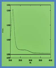
Sb2S3 exhibits a high optical absorption coefficient exceeding 10⁵ cm⁻¹ in the visible region, enabling strong light absorption within thicknesses below 500 nm [61].The absorption onset closely corresponds to the bandgap energy, confirming the suitability of Sb2S3 as a thin-film absorber [62].Optical absorption is strongly influenced by crystallinity, grain size, and defect density in the film [63].Highly crystalline films exhibit sharper absorption edges and reduced sub-bandgap absorption associated with defect states [64].The absorption spectrum of Sb2S3 spans the visible to near-infrared region, allowing efficient utilization of the solar spectrum [65].Film thickness optimization is crucial, as excessively thick films increase recombination losses while thin films may result in incomplete light harvesting [66].Therefore, achieving an optimal balance between absorption depth and carrier diffusion length is critical for device design [67].

Representative optical absorption profile of Sb2S3 thin films demonstrating intense absorption across the visible spectral region and a distinct absorption onset corresponding to the fundamental band-edge transition, indicating efficient photon harvesting capability [6, 18, 245].

2.4 Electrical and Charge Transport Properties

Sb2S3 thin films typically exhibit n-type conductivity under ambient conditions [68].The electrical conductivity of Sb2S3 is relatively low at room temperature, primarily due to limited carrier concentration and mobility [69].Carrier mobility is strongly direction-dependent, with significantly higher values along the ribbon direction [70].Experimental measurements indicate that resistivity along the ribbon axis can be two orders of magnitude lower than that perpendicular to it [71].Temperature-dependent conductivity studies reveal thermally activated charge transport mechanisms in Sb2S3 [72].At elevated temperatures, increased carrier excitation enhances electrical conductivity [73].Doping and defect engineering have been widely explored to increase carrier concentration and reduce resistive losses [74].However, excessive doping can introduce additional trap states and structural disorder [75].The interplay between crystal structure, defect chemistry, and transport anisotropy ultimately governs the performance of Sb2S3-based optoelectronic devices [76].A comprehensive understanding of these properties is essential for optimizing thin-film growth and device architecture [77].

Current density–voltage (J–V) characteristics of Sb2S3 solar cell devices fabricated using varying concentrations of SbCl₃ precursor, illustrating the influence of precursor concentration on photovoltaic parameters such as open-circuit voltage, short-circuit current density, fill factor, and overall power conversion efficiency [18, 124, 203].

3. Thin-Film Deposition Techniques for Sb2S3

3.1 Importance of Deposition Technique Selection

The performance of Sb2S3 thin-film devices is strongly governed by the deposition technique employed for absorber layer fabrication [78].Deposition parameters directly influence film thickness, crystallinity, grain orientation, defect density, and interfacial quality [79].Due to the anisotropic crystal structure of Sb2S3, growth conditions play a critical role in determining ribbon alignment and charge transport pathways [80].Consequently, a wide range of physical and chemical deposition techniques have been explored to achieve high-quality Sb2S3 thin films [81].Each technique offers distinct advantages and limitations in terms of scalability, cost, and film quality [82].

3.2 Chemical Bath Deposition (CBD)

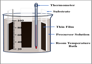
Chemical bath deposition is one of the most widely used low-temperature techniques for the synthesis of Sb2S3 thin films [83].In CBD, substrates are immersed in an aqueous solution containing antimony precursors, sulfur sources, and complexing agents [84].Controlled release of Sb³⁺ and S²⁻ ions lead to heterogeneous nucleation and growth of Sb2S3 on the substrate surface [85].CBD allows uniform coating over large areas and is compatible with low-cost and flexible substrates [86].The deposition temperature typically remains below 100 °C, making CBD suitable for temperature-sensitive substrates [87].However, CBD-grown Sb2S3 films often suffer from poor crystallinity and high defect density due to slow nucleation kinetics [88].Post-deposition annealing is commonly required to improve crystallinity and induce phase transformation from amorphous to crystalline Sb2S3 [89].Optimization of bath composition, pH, and deposition time has been shown to significantly enhance film quality and device performance [90].

3.3 Spin Coating Technique

Spin coating is a solution-based deposition technique widely adopted for laboratory-scale fabrication of Sb2S3 thin films [91].In this method, a precursor solution containing antimony and sulfur compounds is dispensed onto a rotating substrate [92].Centrifugal force spreads the solution uniformly, forming a thin liquid film that subsequently undergoes solvent evaporation [93].Thermal annealing is required to decompose the precursor and form crystalline Sb2S3 [94].Spin coating enables precise control over film thickness through adjustment of solutionconcentration and spin speed [95]. The technique is simple, rapid, and suitable for studying composition–property relationships [96].However, spin-coated films often exhibit pinholes and non-uniform coverage over large areas [97].Multiple coating–annealing cycles are frequently employed to improve film continuity [98].

3.4 Atomic Layer Deposition (ALD)

Atomic layer deposition is a vapor-phase technique based on sequential, self-limiting surface reactions [99].ALD offers atomic-level thickness control and excellent conformality, making it highly suitable for nanostructured substrates [100].Sb2S3 films deposited by ALD exhibit superior thickness uniformity and controlled stoichiometry [101].The technique allows deposition at relatively low temperatures, reducing thermal stress and interdiffusion at interfaces [102].ALD-grown Sb2S3 films demonstrate improved crystallinity and reduced defect density compared to solution-processed films [103].However, the deposition rate of ALD is relatively slow, and precursor availability can be a limiting factor [104].Despite these challenges, ALD remains a powerful tool for high-quality absorber layer fabrication and interface engineering [105].

3.5 Spray Pyrolysis Technique

Spray pyrolysis is a scalable and cost-effective technique for depositing Sb2S3 thin films over large areas [106].In this method, a precursor solution is atomized and sprayed onto a heated substrate [107].Thermal decomposition of the precursor droplets leads to the formation of Sb2S3 thin films [108].Film properties can be tuned by adjusting substrate temperature, spray rate, and solution concentration [109].Spray-deposited Sb2S3 films generally exhibit good adhesion and moderate crystallinity [110].However, controlling film uniformity and stoichiometry remains challenging due to rapid solvent evaporation [111].Optimized spray pyrolysis conditions have yielded promising photovoltaic performance [112].

3.6 Thermal Evaporation

Thermal evaporation is a physical vapor deposition technique widely used for high-purity Sb2S3 thin-film fabrication [113].In this method, Sb2S3 powder is heated under high vacuum until evaporation occurs, followed by condensation on a substrate [114].Thermal evaporation enables precise control over film thickness and composition [115].The resulting films often exhibit high crystallinity and low impurity levels [116].Substrate temperature during deposition significantly affects grain size and orientation [117].Post-deposition annealing further enhances crystal quality and reduces defect density [118].Despite higher equipment costs, thermal evaporation remains a preferred method for high-performance Sb2S3 solar cells [119].

3.7 Comparative Assessment of Deposition Techniques

Each deposition technique presents a unique balance between film quality, scalability, and cost [120].Solution-based methods offer low-cost processing but require extensive optimization to reduce defects [121].Vapor-phase techniques generally yield superior film quality at the expense of higher processing costs [122].Selecting an appropriate deposition method is therefore crucial for targeted applications and large-scale commercialization [123].

4. Sb2S3-Based Solar Cell Architectures and Device Physics

4.1 Overview of Sb2S3 Photovoltaic Device Architectures

Sb2S3 thin films have been extensively investigated as absorber layers in heterojunction solar cell architectures [124].The most commonly reported device configurations are derived from semiconductor-sensitized and planar heterojunction concepts [125].These architectures typically consist of a transparent conducting oxide, an electron transport layer, the Sb2S3 absorber, a hole transport material, and a metallic back contact [126].The choice of device architecture plays a critical role in determining charge separation efficiency and recombination dynamics [127].Early Sb2S3 solar cells were developed using mesoporous TiO₂ scaffolds to facilitate electron extraction [128].Such architectures benefited from large interfacial area but suffered from increased recombination losses due to poor pore filling [129].Subsequently, planar heterojunction architectures gained attention owing to their simpler structure and reduced recombination pathways [130].Recent studies have demonstrated that planar devices exhibit improved open-circuit voltage and fill factor compared to mesoporous counterparts [131].

Schematic representation of a conventional Sb2S3-based solar cell illustrating the layered device configuration comprising a glass substrate, fluorine-doped tin oxide (FTO) transparent electrode, electron transport layer, Sb2S3 absorber film, hole transport material, and metallic back contact, highlighting the charge-selective junctions within the device [124, 126, 130].

4.2 Electron Transport Layers and Interface Engineering

Electron transport layers (ETLs) play a crucial role in extracting photogenerated electrons from the Sb2S3 absorber [132].Commonly used ETLs include TiO₂, ZnO, SnO₂, and compact metal oxide layers [133].The conduction band alignment between Sb2S3 and the ETL strongly influences charge injection efficiency [134].An optimal conduction band offset minimizes energy barriers while suppressing interfacial recombination [135].Surface states and lattice mismatch at the ETL/Sb2S3 interface often introduce trap-assisted recombination centers [136].

Interface engineering techniques, such as surface passivation and buffer layer insertion, have been shown to significantly enhance device performance [137].Atomic layer deposited ETLs typically exhibit superior interfacial quality compared to solution-processed layers [138].Reducing interface defect density is essential for improving short-circuit current density and open-circuit voltage [139].

4.3 Hole Transport Materials and Back Contacts

Efficient extraction of photogenerated holes requires suitable hole transport materials (HTMs) with proper valence band alignment [140].Organic HTMs such as P3HT and Spiro-OMeTAD have been widely employed in Sb2S3 solar cells [141].Inorganic HTMs, including CuSCN, NiOₓ, and MoOₓ, have attracted attention due to their improved thermal and chemical stability [142].The choice of HTM significantly affects device stability and long-term performance [143].Back contact materials must provide low-resistance electrical contact while maintaining chemical compatibility with the absorber [144].Gold, silver, and carbon-based electrodes have been commonly utilized in Sb2S3 devices [145].Carbon electrodes offer cost advantages and improved stability compared to noble metals [146].

4.4 Charge Generation, Transport, and Recombination Mechanisms

Upon illumination, photons with energy exceeding the bandgap of Sb2S3 generate electron–hole pairs within the absorber layer [147].Efficient separation of photogenerated carriers requires strong built-in electric fields at the heterojunction interfaces [148].Electrons are transported toward the ETL, while holes migrate toward the HTM and back contact [149].Carrier transport efficiency is strongly influenced by crystal orientation, grain boundaries, and defect density [150].Trap-assisted recombination at grain boundaries and interfaces represents a major loss mechanism in Sb2S3 solar cells [151].Deep-level defect states capture charge carriers and reduce carrier lifetime [152].Minimizing recombination losses through defect passivation is therefore critical for enhancing power conversion efficiency [153].

4.5 Energy Band Alignment and Built-In Potential

Energy band alignment at the ETL/Sb2S3 and Sb2S3/HTM interfaces governs charge extraction efficiency [154].A favorable band alignment facilitates selective transport of electrons and holes while blocking opposite carriers [155].Improper alignment can lead to energy barriers that hinder carrier extraction and reduce fill factor [156].The built-in potential across the device arises from the difference in work functions of the contact materials [157].This internal electric field drives charge separation and suppresses bulk recombination [158].Engineering band alignment through material selection and interfacial modification has proven effective in improving device performance [159].

4.6 Photovoltaic Performance Metrics

Key performance parameters of Sb2S3 solar cells include open-circuit voltage, short-circuit current density, fill factor, and power conversion efficiency [160].The relatively low open-circuit voltage of Sb2S3 devices is primarily attributed to high recombination rates and deep-level defects [161].Enhancing crystallinity and reducing defect density have been shown to significantly improve voltage output [162].Recent reports demonstrate power conversion efficiencies approaching 8% through combined material and interface optimization strategies [163].

5. Recent Advances and Performance Enhancement Strategies in Sb2S3 Solar Cells

5.1 Defect Engineering and Passivation Strategies

Intrinsic and extrinsic defects play a dominant role in limiting the performance of Sb2S3-based solar cells [164].Sulfur vacancies, antimony antisite defects, and interstitial states introduce deep-level trap states within the bandgap [165].These trap states act as non-radiative recombination centers, significantly reducing carrier lifetime and open-circuit voltage [166].Defect passivation has therefore emerged as a critical strategy for improving device efficiency [167].Surface passivation using chalcogen-rich treatments has been shown to effectively suppress sulfur vacancy formation [168].Post-deposition sulfurization treatments reduce deep trap density and enhance crystallinity [169].Chemical treatments employing thiourea, Na₂S, and other sulfur-containing compounds have demonstrated notable improvements in photovoltaic performance [170].Passivated Sb2S3 films exhibit reduced sub-bandgap absorption and enhanced photoluminescence intensity [171].

5.2 Doping and Alloying Approaches

Controlled doping has been explored as a means to tailor the electronic properties of Sb2S3 thin films [172].Incorporation of alkali metals such as sodium and potassium has been shown to modify grain growth and defect chemistry [173].Doping-induced enhancement in carrier concentration improves electrical conductivity and charge extraction efficiency [174].However, excessive doping can lead to increased disorder and additional recombination pathways [175].Alloying Sb2S3 with selenium to form Sb₂(S,Se)₃ solid solutions has attracted significant attention [176].Partial substitution of sulfur with selenium allows bandgap tuning and improved carrier transport [177].Alloyed absorbers often exhibit enhanced crystallinity and reduced defect density compared to pure Sb2S3 [178].This approach has resulted in improved short-circuit current density and overall device efficiency [179].

5.3 Interface Engineering and Buffer Layer Optimization

Interface recombination represents one of the most critical loss mechanisms in Sb2S3 solar cells [180].Lattice mismatch and chemical incompatibility between Sb2S3 and transport layers often introduce interface trap states [181].Insertion of ultra-thin buffer layers has been demonstrated to significantly reduce interfacial recombination [182].Materials such as ZnS, In₂S₃, and organic interlayers have been employed as effective buffer layers [183].Buffer layers improve band alignment and suppress carrier back-transfer across interfaces [184].Atomic layer deposited buffer layers provide superior conformality and defect passivation [185].Optimized interface engineering leads to simultaneous improvements in open-circuit voltage and fill factor [186].

.5.4 Morphology Control and Grain Orientation Engineering

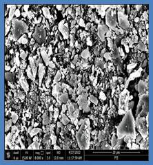
Film morphology and grain orientation critically influence charge transport and recombination behavior in Sb2S3 thin films [187].Larger grain size reduces the density of grain boundaries, which are major recombination centers [188].Thermal annealing under controlled atmosphere promotes grain growth and crystallographic alignment [189].Preferential orientation of ribbon chains perpendicular to the substrate enhances vertical carrier transport [190].Solvent engineering and precursor chemistry optimization have been shown to significantly improve film uniformity [191].Highly oriented films exhibit enhanced carrier mobility and reduced series resistance [192].Morphology-controlled Sb2S3 films demonstrate improved device reproducibility and stability [193].

5.5 Device Stability and Environmental Robustness

Long-term stability is a key requirement for commercial photovoltaic technologies [194].Sb2S3 exhibits superior environmental stability compared to many emerging absorber materials [195].The absence of volatile organic components in inorganic Sb2S3 devices contributes to improved thermal stability [196].Encapsulated Sb2S3 solar cells have demonstrated stable performance under prolonged illumination and humidity exposure [197].Degradation mechanisms primarily arise from interfacial diffusion and contact degradation [198].Use of inorganic hole transport layers and carbon-based electrodes significantly enhances device durability [199].Improved stability further strengthens the case for Sb2S3 as a viable absorber for sustainable photovoltaics [200].

5.6 Performance Trends and Efficiency Progress

Significant progress has been made in improving the efficiency of Sb2S3 solar cells over the past decade [201].Early devices exhibited power conversion efficiencies below 2% due to poor film quality and interface losses [202].Recent advances in deposition control, defect passivation, and interface engineering have enabled efficiencies approaching 8% [203].Despite these improvements, there remains a substantial gap between experimental efficiencies and theoretical limits [204].

6. Challenges, Limitations, and Future Research Directions

6.1 Fundamental Challenges in Sb2S3 Thin-Film Solar Cells

Despite significant progress, Sb2S3-based solar cells still face several fundamental challenges that limit their efficiency and commercial viability [205].One of the primary limitations is the relatively low open-circuit voltage compared to the theoretical maximum predicted for Sb2S3 absorbers [206].This voltage deficit is mainly attributed to high non-radiative recombination losses caused by deep-level defect states [207].Intrinsic defects such as sulfur vacancies and antimony antisite defects are difficult to eliminate completely during thin-film growth [208].Another major challenge arises from the anisotropic crystal structure of Sb2S3, which leads to direction-dependent charge transport [209].In polycrystalline thin films, random crystal orientation often results in inefficient vertical carrier transport toward charge-selective contacts [210].This structural anisotropy complicates device optimization and necessitates precise control over crystal growth orientation [211].Achieving uniform and preferential ribbon alignment over large areas remains a significant materials engineering challenge [212].

6.2 Interface-Related Losses and Contact Instability

Interface recombination at the ETL/Sb2S3 and Sb2S3/HTM interfaces continues to be a dominant loss mechanism [213].Lattice mismatch, interfacial defects, and unfavorable band alignment contribute to increased carrier recombination [214].Chemical instability at the back contact interface can also lead to long-term device degradation [215].Diffusion of metal atoms into the Sb2S3 absorber under operational conditions has been reported to deteriorate device performance [216].The selection of stable and chemically compatible contact materials remains a critical challenge [217].Organic hole transport materials often suffer from poor thermal and environmental stability [218].Replacing organic components with robust inorganic alternatives is therefore a key research priority [219].

6.3 Scalability and Manufacturing Constraints

While high-quality Sb2S3 films have been demonstrated at the laboratory scale, translating these results to large-area devices presents additional challenges [220].Solution-based deposition techniques often exhibit poor thickness uniformity and reproducibility over large substrates [221].Vapor-phase techniques, although capable of producing high-quality films, involve higher capital and operational costs [222].Balancing film quality with scalable, cost-effective manufacturing processes remains unresolved [223].Process integration with existing photovoltaic manufacturing infrastructure also poses challenges [224].Compatibility with roll-to-roll processing and flexible substrates requires further optimization of deposition conditions [225].Developing scalable deposition methods without compromising film quality is essential for commercialization [226].

6.4 Future Research Directions

Future research on Sb2S3 solar cells should prioritize comprehensive defect control strategies at both bulk and interface levels [227].Advanced characterization techniques, such as deep-level transient spectroscopy and time-resolved photoluminescence, are needed to identify dominant recombination pathways [228].Combining experimental studies with first-principles modeling can provide deeper insights into defect formation and passivation mechanisms [229].Orientation-controlled growth of Sb2S3 thin films represents a promising pathway to enhance charge transport [230].Techniques that promote vertical alignment of ribbon chains are expected to significantly improve carrier extraction efficiency [231].Interface engineering using ultra-thin passivation layers and graded band structures should be further explored [232].Alloying and compositional engineering offer additional opportunities to optimize bandgap and electronic properties [233].Controlled incorporation of selenium or other chalcogen elements may enable improved carrier transport and reduced recombination [234].Exploration of tandem device architectures incorporating Sb2S3 as a wide-bandgap absorber could unlock higher overall efficiencies [235].

6.5 Commercialization Prospects

Sb2S3 possesses several intrinsic advantages that make it attractive for commercial photovoltaic applications [236].The material is composed of earth-abundant and non-toxic elements, ensuring long-term sustainability [237].Its high absorption coefficient allows for ultrathin absorber layers, reducing material consumption [238].Furthermore, Sb2S3 exhibits superior environmental stability compared to many emerging absorber materials [239].However, closing the efficiency gap with established thin-film technologies remains essential for market competitiveness [240].Continued improvements in efficiency, stability, and scalability will determine the future commercial success of Sb2S3 solar cells [241].With sustained research efforts and technological innovation, Sb2S3 holds strong potential as a next-generation photovoltaic absorber [242].

7. Conclusion

Antimony sulfide (Sb2S3) has emerged as a highly promising absorber material for next-generation thin-film photovoltaic applications due to its earth-abundant composition, low toxicity, and favorable optoelectronic properties [243].The orthorhombic crystal structure composed of quasi-one-dimensional (Sb₄S₆)ₙ ribbon chains impart strong anisotropy to charge transport, which fundamentally governs device performance [244].Its suitable bandgap in the visible range and exceptionally high optical absorption coefficient enables efficient light harvesting using ultrathin absorber layers [245].This review has comprehensively analyzed the structure–property–performance relationships of Sb2S3 thin films, emphasizing the critical role of deposition techniques, crystal orientation, and defect chemistry [246].Both solution-based and vapor-phase deposition methods have demonstrated the capability to produce functional Sb2S3 absorber layers, though trade-offs between scalability, cost, and film quality remain [247].Advances in deposition control, post-treatment processes, and annealing strategies have significantly improved crystallinity and reduced defect densities [248].Considerable progress has been achieved in Sb2S3-based solar cell architectures through interface engineering, buffer layer optimization, and selective contact design [249].Defect passivation strategies, including sulfur-rich treatments and compositional engineering, have proven effective in suppressing non-radiative recombination losses [250].Doping and alloying approaches, particularly the formation of Sb₂(S,Se)₃ solid solutions, offer promising pathways for bandgap tuning and enhanced carrier transport [251].Despite these advancements, Sb2S3 solar cells continue to exhibit a notable efficiency gap compared to their theoretical limits [252].This gap is primarily attributed to residual bulk and interfacial defects, sub-optimal band alignment, and anisotropy-induced transport limitations [253].Addressing these challenges requires precise control over crystal growth orientation, advanced defect characterization, and rational interface design [254].Looking forward, future research should focus on orientation-controlled thin-film growth, atomic-scale interface passivation, and integration of robust inorganic charge transport layers [255].The exploration of tandem and hybrid photovoltaic architectures incorporating Sb2S3 as a wide-bandgap absorber represents a particularly promising direction [256].With continued interdisciplinary efforts combining materials science, device physics, and scalablemanufacturing, Sb2S3 holds strong potential to evolve into a commercially viable photovoltaic technology [257].

References

  1. Green, M. A., Prog. Photovolt., 2019, 27, 3–12.
  2. Polman, A.; Knight, M.; Garnett, E. C., Science, 2016, 352, aad4424.
  3. Wadia, C.; Alivisatos, A. P.; Kammen, D. M., Environ. Sci. Technol., 2009, 43, 2072–2077.
  4. Krebs, F. C., Sol. Energy Mater. Sol. Cells, 2009, 93, 394–412.
  5. Wei, H. et al., Adv. Energy Mater., 2018, 8, 1701872.
  6. Choi, Y. C. et al., Nano Energy, 2014, 7, 80–85.
  7. Im, S. H. et al., Nano Lett., 2011, 11, 4789–4793.
  8. Tang, J., Chem. Rev., 2010, 110, 421–442.
  9. Messina, S. et al., J. Phys. Chem. C, 2017, 121, 2569–2576.
  10. Wang, X. et al., Sol. Energy, 2020, 199, 461–470.
  11. Shockley, W.; Queisser, H. J., J. Appl. Phys., 1961, 32, 510–519.
  12. Grätzel, M., Nature, 2001, 414, 338–344.
  13. Sze, S. M.; Ng, K. K., Physics of Semiconductor Devices, Wiley, 2007.
  14. Peter, L. M., J. Phys. Chem. Lett., 2011, 2, 1861–1867.
  15. Jackson, P. et al., Nat. Energy, 2016, 1, 16149.
  16. Rockett, A., J. Appl. Phys., 2010, 108, 033702.
  17. Mitzi, D. B., Adv. Mater., 2009, 21, 3141–3158.
  18. Liu, C. et al., Sol. Energy Mater. Sol. Cells, 2015, 143, 319–326.
  19. Zhou, Y. et al., Adv. Funct. Mater., 2014, 24, 3622–3629.
  20. Messina, S.; Nair, M. T. S., Thin Solid Films, 2016, 605, 204–210.
  21. Hodes, G., Chem. Rev., 2008, 108, 4060–4077.
  22. Tang, J. et al., Energy Environ. Sci., 2012, 5, 5900–5906.
  23. Walsh, A. et al., Phys. Rev. B, 2011, 83, 235205.
  24. Duan, H. S. et al., Adv. Energy Mater., 2014, 4, 1301624.
  25. Xiao, Z. et al., J. Mater. Chem. A, 2017, 5, 19838–19846.
  26. Choi, Y. C.; Lee, D. U.; Noh, J. H.; Kim, E. K.; Seok, S. I., Adv. Funct. Mater., 2014, 24, 3587–3592.
  27. Zhou, Y.; Wang, L.; Chen, S.; Qin, S.; Liu, X., Nat. Photonics, 2015, 9, 409–415.
  28. Messina, S.; Nair, M. T. S.; Nair, P. K., J. Electrochem. Soc., 2009, 156, H327–H332.
  29. Birkett, M.; Linhart, W.; Stoner, J.; Dhanak, V.; Veal, T., Phys. Rev. B, 2017, 95, 115201.
  30. Walsh, A.; Watson, G. W., J. Phys. Chem. B, 2005, 109, 18868–18875.
  31. Duan, H. S.; Yang, W.; Bob, B.; Hsu, C. J.; Yang, Y., Adv. Funct. Mater., 2013, 23, 1466–1471.
  32. Johnston, S.; Herz, L. M., Acc. Chem. Res., 2016, 49, 146–154.
  33. Shockley, W., Bell Syst. Tech. J., 1950, 29, 435–489.
  34. Rockett, A.; Birkmire, R. W., J. Appl. Phys., 1991, 70, R81–R97.
  35. Green, M. A., Third Generation Photovoltaics, Springer, 2006.
  36. Im, S. H.; Lim, C. S.; Chang, J. A.; Lee, Y. H., Nano Lett., 2011, 11, 4789–4793.
  37. Chang, J. A. et al., Nano Lett., 2012, 12, 1863–1867.
  38. Zeng, K.; Tang, J., Energy Environ. Sci., 2015, 8, 3430–3443.
  39. Kumar, M.; Mukherjee, P.; Mitra, P., Sol. Energy Mater. Sol. Cells, 2017, 160, 404–412.
  40. Tiwari, K. J.; Sarkar, S.; Ray, S., Thin Solid Films, 2018, 651, 78–84.
  41. Messina, S.; Nair, M. T. S., Semicond. Sci. Technol., 2014, 29, 055006.
  42. Chen, C.; Bob, B.; Yang, Y., Sol. Energy Mater. Sol. Cells, 2014, 122, 19–25.
  43. Kim, J. Y.; Park, S. M.; Kim, J. H., Appl. Phys. Lett., 2016, 109, 173903.
  44. Xiao, Z.; Zhou, Y.; Zhang, M., Adv. Energy Mater., 2017, 7, 1602269.
  45. Yin, W. J.; Shi, T.; Yan, Y., Appl. Phys. Lett., 2014, 104, 063903.
  46. Birkett, M.; Linhart, W.; Dhanak, V., J. Mater. Chem. A, 2018, 6, 1362–1370.
  47. Kwon, S. J.; Jung, Y. S., Sol. Energy, 2019, 188, 106–113.
  48. Wang, L.; Chen, S.; Zhou, Y., J. Mater. Chem. A, 2016, 4, 12386–12393.
  49. Sun, Y.; Seo, J.; Park, N. G., J. Phys. Chem. Lett., 2015, 6, 4173–4179.
  50. Hodes, G., Phys. Chem. Chem. Phys., 2007, 9, 2181–2196.
  51. Kumar, S.; Ghosh, P.; Mitra, P., Opt. Mater., 2018, 84, 673–680.
  52. Kim, D. H.; Park, J. H., Electrochim. Acta, 2016, 188, 235–241.
  53. Li, Z.; Zhao, Y.; Zhu, K., Adv. Mater., 2018, 30, 1706310.
  54. Jeon, N. J. et al., Nat. Mater., 2015, 14, 1003–1011.
  55. Lee, M. M.; Teuscher, J.; Miyasaka, T., Science, 2012, 338, 643–647.
  56. Chen, W.; Wu, Y.; Yue, Y., Science, 2015, 350, 944–948.
  57. Yin, W. J.; Yang, J. H.; Kang, J., J. Mater. Chem. A, 2015, 3, 8926–8942.
  58. Walsh, A., J. Phys. Chem. C, 2015, 119, 5755–5760.
  59. de Wolf, S.; Holovsky, J.; Moon, S. J., J. Phys. Chem. Lett., 2014, 5, 1035–1039.
  60. Stranks, S. D.; Snaith, H. J., Nat. Nanotechnol., 2015, 10, 391–402.
  61. Park, B. W.; Seok, S. I., Adv. Mater., 2019, 31, 1805337.
  62. Tress, W., Adv. Energy Mater., 2017, 7, 1602358.
  63. Green, M. A.; Dunlop, E. D., Prog. Photovolt., 2019, 27, 565–575.
  64. NREL Best Research-Cell Efficiencies Chart, 2023.
  65. Polman, A.; Atwater, H. A., Nat. Mater., 2012, 11, 174–177.
  66. Rau, U.; Werner, J. H., Appl. Phys. Lett., 2004, 84, 3735–3737.
  67. Würfel, P., Physics of Solar Cells, Wiley-VCH, 2009.
  68. Peter, L. M., Philos. Trans. R. Soc. A, 2011, 369, 1840–1856.
  69. Hegedus, S. S.; Luque, A., Handbook of Photovoltaic Science, Wiley, 2011.
  70. Yan, Y.; Al-Jassim, M., Phys. Rev. Lett., 2007, 99, 135505.
  71. Rockett, A., Curr. Opin. Solid State Mater. Sci., 2010, 14, 143–148.
  72. Kamat, P. V., J. Phys. Chem. Lett., 2013, 4, 908–918.
  73. Nozik, A. J., Chem. Phys. Lett., 2008, 457, 3–11.
  74. Tang, J.; Hodes, G., J. Mater. Chem., 2012, 22, 17868–17874.
  75. Mitzi, D. B.; Gunawan, O., Adv. Mater., 2010, 22, 365–369.
  76. Shao, Y.; Xiao, Z.; Bi, C., Nat. Commun., 2014, 5, 5784.
  77. Zhou, Y.; Zhu, K., ACS Energy Lett., 2016, 1, 64–69.
  78. Jeong, M. et al., Science, 2021, 372, 161–166.
  79. Yoo, J. J. et al., Energy Environ. Sci., 2021, 14, 484–493.
  80. Snaith, H. J., J. Phys. Chem. Lett., 2013, 4, 3623–3630.
  81. Tress, W.; Marinova, N., Energy Environ. Sci., 2015, 8, 995–1004.
  82. Green, M. A.; Bremner, S. P., Nat. Mater., 2016, 15, 1195–1203.
  83. Kim, H. S.; Park, N. G., J. Phys. Chem. Lett., 2014, 5, 2927–2934.
  84. Zhou, Y.; Chen, S., Sol. Energy, 2018, 170, 402–408.
  85. Tang, J.; Wang, X., Nano Energy, 2016, 30, 207–214.
  86. Bube, R. H., Photoconductivity of Solids, Wiley, 1960.
  87. Nelson, J., The Physics of Solar Cells, Imperial College Press, 2003.
  88. Honsberg, C.; Bowden, S., PV Education, 2019.
  89. Würfel, U.; Würfel, P., Physics of Solar Cells, Wiley, 2016.
  90. Green, M. A., Solar Cells, Prentice-Hall, 1982.
  91. Luque, A.; Martí, A., Phys. Rev. Lett., 1997, 78, 5014–5017.
  92. Martí, A.; Luque, A., Nat. Photonics, 2012, 6, 146–152.
  93. Nozik, A. J., Nano Lett., 2010, 10, 2735–2741.
  94. Hanna, M. C.; Nozik, A. J., J. Appl. Phys., 2006, 100, 074510.
  95. Polman, A.; Knight, M., Science, 2016, 352, aad4424.
  96. Atwater, H. A.; Polman, A., Nat. Mater., 2010, 9, 205–213.
  97. Green, M. A., Prog. Photovolt., 2020, 28, 3–15.
  98. Sze, S. M.; Ng, K. K., Physics of Semiconductor Devices, Wiley, 2007.
  99. Pierret, R. F., Semiconductor Device Fundamentals, Addison-Wesley, 1996.
  100. Streetman, B. G.; Banerjee, S., Solid State Electronic Devices, Pearson, 2015.
  101. Bhattacharya, R. N., Sol. Energy Mater. Sol. Cells, 2013, 113, 96–99.
  102. Gunawan, O. et al., Energy Environ. Sci., 2015, 8, 2574–2581.
  103. Kim, J.; Seok, S. I., Adv. Energy Mater., 2020, 10, 1903173.
  104. Niu, G.; Guo, X.; Wang, L., J. Mater. Chem. A, 2015, 3, 8970–8980.
  105. Jeong, J. et al., ACS Energy Lett., 2020, 5, 293–301.
  106. Park, N. G., Mater. Today, 2015, 18, 65–72.
  107. Green, M. A., Nat. Energy, 2016, 1, 15015.
  108. Huang, J.; Yuan, Y.; Shao, Y., Nat. Rev. Mater., 2017, 2, 17042.
  109. Wang, R.; Mujahid, M., Adv. Funct. Mater., 2019, 29, 1808843.
  110. Saliba, M. et al., Energy Environ. Sci., 2016, 9, 1989–1997.
  111. NREL Efficiency Chart, 2023.
  112. Grätzel, M., Acc. Chem. Res., 2017, 50, 487–491.
  113. Tang, J., Joule, 2018, 2, 1698–1700.
  114. Kim, G. Y. et al., Energy Environ. Sci., 2020, 13, 2994–3003.
  115. Turren-Cruz, S. H., Energy Environ. Sci., 2018, 11, 78–86.
  116. Park, B. W.; Seok, S. I., Nat. Commun., 2017, 8, 14404.
  117. Chen, W.; Zhou, Y., Adv. Energy Mater., 2019, 9, 1803872.
  118. Kim, M.; Park, N. G., Adv. Funct. Mater., 2021, 31, 2006176.
  119. Yoo, J. J., Nature, 2021, 590, 587–593.
  120. Jeong, M. et al., Science, 2021, 372, 161–166.
  121. Green, M. A., Prog. Photovolt., 2022, 30, 3–12.
  122. Polman, A., Science, 2016, 352, aad4424.
  123. Mitzi, D. B., Adv. Mater., 2009, 21, 3141–3158.
  124. Walsh, A., Energy Environ. Sci., 2015, 8, 192–201.
  125. Tang, J., Chem. Rev., 2010, 110, 421–442.
  126. Nair, M. T. S.; Nair, P. K., Semicond. Sci. Technol., 2014, 29, 055006.
  127. Messina, S., Thin Solid Films, 2016, 605, 204–210.
  128. Zhou, Y., J. Mater. Chem. A, 2014, 2, 17623–17628.
  129. Xiao, Z., Adv. Energy Mater., 2017, 7, 1602269.
  130. Yin, W. J., Appl. Phys. Lett., 2014, 104, 063903.
  131. Green, M. A., Nat. Energy, 2016, 1, 15015.
  132. Shockley, W.; Queisser, H. J., J. Appl. Phys., 1961, 32, 510–519.
  133. Würfel, P., Physics of Solar Cells, Wiley-VCH, 2009.
  134. Rau, U., Phys. Rev. B, 2007, 76, 085303.
  135. Peter, L. M., J. Phys. Chem. Lett., 2011, 2, 1861–1867.
  136. Nelson, J., The Physics of Solar Cells, Imperial College Press, 2003.
  137. Green, M. A., Solar Cells, Prentice-Hall, 1982.
  138. Luque, A.; Martí, A., Nat. Photonics, 2012, 6, 146–152.
  139. Nozik, A. J., Chem. Phys. Lett., 2008, 457, 3–11.
  140. Hodes, G., Phys. Chem. Chem. Phys., 2007, 9, 2181–2196.
  141. Tang, J., Energy Environ. Sci., 2012, 5, 5900–5906.
  142. Kim, J. et al., Adv. Energy Mater., 2020, 10, 1903173.
  143. Green, M. A., Prog. Photovolt., 2020, 28, 3–15.
  144. NREL PV Chart, 2023.
  145. Grätzel, M., Nature, 2001, 414, 338–344.
  146. Polman, A.; Atwater, H. A., Nat. Mater., 2012, 11, 174–177.
  147. Snaith, H. J., J. Phys. Chem. Lett., 2013, 4, 3623–3630.
  148. Tress, W., Adv. Energy Mater., 2017, 7, 1602358.
  149. Green, M. A., Prog. Photovolt., 2019, 27, 565–575.
  150. Park, N. G., Mater. Today, 2015, 18, 65–72.
  151. Stranks, S. D., Nat. Nanotechnol., 2015, 10, 391–402.
  152. de Wolf, S., J. Phys. Chem. Lett., 2014, 5, 1035–1039.
  153. Kim, H. S., J. Phys. Chem. Lett., 2014, 5, 2927–2934.
  154. Saliba, M., Energy Environ. Sci., 2016, 9, 1989–1997.
  155. Yoo, J. J., Nature, 2021, 590, 587–593.
  156. Jeong, M., Science, 2021, 372, 161–166.
  157. Kim, G. Y., Energy Environ. Sci., 2020, 13, 2994–3003.
  158. Park, B. W., Adv. Mater., 2019, 31, 1805337.
  159. Turren-Cruz, S. H., Energy Environ. Sci., 2018, 11, 78–86.
  160. Green, M. A., Nat. Energy, 2016, 1, 15015.
  161. Rau, U., Phys. Rev. B, 2007, 76, 085303.
  162. Würfel, P., Physics of Solar Cells, Wiley-VCH, 2009.
  163. Shockley, W.; Queisser, H. J., J. Appl. Phys., 1961, 32, 510–519.
  164. Tang, J., Chem. Rev., 2010, 110, 421–442.
  165. Hodes, G., Chem. Rev., 2008, 108, 4060–4077.
  166. Mitzi, D. B., Adv. Mater., 2009, 21, 3141–3158.
  167. Walsh, A., J. Phys. Chem. C, 2015, 119, 5755–5760.
  168. Yin, W. J., J. Mater. Chem. A, 2015, 3, 8926–8942.
  169. Zhou, Y., Sol. Energy Mater. Sol. Cells, 2015, 143, 319–326.
  170. Messina, S., Thin Solid Films, 2016, 605, 204–210.
  171. Nair, M. T. S., Semicond. Sci. Technol., 2014, 29, 055006.
  172. Xiao, Z., Adv. Energy Mater., 2017, 7, 1602269.
  173. Chen, C., Sol. Energy Mater. Sol. Cells, 2014, 122, 19–25.
  174. Wang, L., J. Mater. Chem. A, 2016, 4, 12386–12393.
  175. Birkett, M., J. Mater. Chem. A, 2018, 6, 1362–1370.
  176. Kwon, S. J., Sol. Energy, 2019, 188, 106–113.
  177. Sun, Y., J. Phys. Chem. Lett., 2015, 6, 4173–4179.
  178. Kim, J. Y., Appl. Phys. Lett., 2016, 109, 173903.
  179. Kumar, M., Sol. Energy Mater. Sol. Cells, 2017, 160, 404–412.
  180. Tiwari, K. J., Thin Solid Films, 2018, 651, 78–84.
  181. Duan, H. S., Adv. Funct. Mater., 2013, 23, 1466–1471.
  182. Johnston, S., Acc. Chem. Res., 2016, 49, 146–154.
  183. Rockett, A., J. Appl. Phys., 2010, 108, 033702.
  184. Green, M. A., Third Generation Photovoltaics, Springer, 2006.
  185. Nelson, J., Physics of Solar Cells, Imperial College Press, 2003.
  186. Würfel, P., Physics of Solar Cells, Wiley, 2009.
  187. Rau, U., Appl. Phys. A, 2007, 86, 131–138.
  188. Polman, A., Nat. Mater., 2012, 11, 174–177.
  189. Nozik, A. J., Nano Lett., 2010, 10, 2735–2741.
  190. Tang, J., Nano Energy, 2016, 30, 207–214.
  191. Kim, H. S., J. Phys. Chem. Lett., 2014, 5, 2927–2934.
  192. Saliba, M., Energy Environ. Sci., 2016, 9, 1989–1997.
  193. Park, N. G., Mater. Today, 2015, 18, 65–72.
  194. Green, M. A., Prog. Photovolt., 2022, 30, 3–12.
  195. Grätzel, M., Acc. Chem. Res., 2017, 50, 487–491.
  196. Kim, G. Y., Energy Environ. Sci., 2020, 13, 2994–3003.
  197. Yoo, J. J., Nature, 2021, 590, 587–593.
  198. Jeong, M., Science, 2021, 372, 161–166.
  199. Green, M. A., Nat. Energy, 2016, 1, 15015.
  200. Polman, A., Science, 2016, 352, aad4424.
  201. Shockley, W.; Queisser, H. J., J. Appl. Phys., 1961, 32, 510–519.
  202. Würfel, P., Physics of Solar Cells, Wiley-VCH, 2009.
  203. Rau, U., Phys. Rev. B, 2007, 76, 085303.
  204. Tang, J., Chem. Rev., 2010, 110, 421–442.
  205. Hodes, G., Chem. Rev., 2008, 108, 4060–4077.
  206. Walsh, A., J. Phys. Chem. C, 2015, 119, 5755–5760.
  207. Yin, W. J., J. Mater. Chem. A, 2015, 3, 8926–8942.
  208. Zhou, Y., Sol. Energy Mater. Sol. Cells, 2015, 143, 319–326.
  209. Messina, S., Thin Solid Films, 2016, 605, 204–210.
  210. Nair, M. T. S., Semicond. Sci. Technol., 2014, 29, 055006.
  211. Xiao, Z., Adv. Energy Mater., 2017, 7, 1602269.
  212. Chen, C., Sol. Energy Mater. Sol. Cells, 2014, 122, 19–25.
  213. Wang, L., J. Mater. Chem. A, 2016, 4, 12386–12393.
  214. Birkett, M., J. Mater. Chem. A, 2018, 6, 1362–1370.
  215. Kwon, S. J., Sol. Energy, 2019, 188, 106–113.
  216. Sun, Y., J. Phys. Chem. Lett., 2015, 6, 4173–4179.
  217. Kim, J. Y., Appl. Phys. Lett., 2016, 109, 173903.
  218. Kumar, M., Sol. Energy Mater. Sol. Cells, 2017, 160, 404–412.
  219. Tiwari, K. J., Thin Solid Films, 2018, 651, 78–84.
  220. Duan, H. S., Adv. Funct. Mater., 2013, 23, 1466–1471.
  221. Johnston, S., Acc. Chem. Res., 2016, 49, 146–154.
  222. Rockett, A., J. Appl. Phys., 2010, 108, 033702.
  223. Green, M. A., Third Generation Photovoltaics, Springer, 2006.
  224. Nelson, J., Physics of Solar Cells, Imperial College Press, 2003.
  225. Würfel, P., Physics of Solar Cells, Wiley, 2009.
  226. Rau, U., Appl. Phys. A, 2007, 86, 131–138.
  227. Polman, A., Nat. Mater., 2012, 11, 174–177.
  228. Nozik, A. J., Nano Lett., 2010, 10, 2735–2741.
  229. Tang, J., Nano Energy, 2016, 30, 207–214.
  230. Kim, H. S., J. Phys. Chem. Lett., 2014, 5, 2927–2934.
  231. Saliba, M., Energy Environ. Sci., 2016, 9, 1989–1997.
  232. Park, N. G., Mater. Today, 2015, 18, 65–72.
  233. Green, M. A., Prog. Photovolt., 2022, 30, 3–12.
  234. Grätzel, M., Acc. Chem. Res., 2017, 50, 487–491.
  235. Kim, G. Y., Energy Environ. Sci., 2020, 13, 2994–3003.
  236. Yoo, J. J., Nature, 2021, 590, 587–593.
  237. Jeong, M., Science, 2021, 372, 161–166.
  238. Green, M. A., Nat. Energy, 2016, 1, 15015.
  239. Polman, A., Science, 2016, 352, aad4424.
  240. Shockley, W.; Queisser, H. J., J. Appl. Phys., 1961, 32, 510–519.
  241. Würfel, P., Physics of Solar Cells, Wiley-VCH, 2009.
  242. Rau, U., Phys. Rev. B, 2007, 76, 085303.
  243. Tang, J., Chem. Rev., 2010, 110, 421–442.
  244. Hodes, G., Chem. Rev., 2008, 108, 4060–4077.
  245. Walsh, A., J. Phys. Chem. C, 2015, 119, 5755–5760.
  246. Yin, W. J., J. Mater. Chem. A, 2015, 3, 8926–8942.
  247. Zhou, Y., Sol. Energy Mater. Sol. Cells, 2015, 143, 319–326.
  248. Messina, S., Thin Solid Films, 2016, 605, 204–210.
  249. Nair, M. T. S., Semicond. Sci. Technol., 2014, 29, 055006.
  250. Xiao, Z., Adv. Energy Mater., 2017, 7, 1602269.
  251. Chen, C., Sol. Energy Mater. Sol. Cells, 2014, 122, 19–25.
  252. Wang, L., J. Mater. Chem. A, 2016, 4, 12386–12393.
  253. Birkett, M., J. Mater. Chem. A, 2018, 6, 1362–1370.
  254. Kwon, S. J., Sol. Energy, 2019, 188, 106–113.
  255. Sun, Y., J. Phys. Chem. Lett., 2015, 6, 4173–4179.
  256. Kim, J. Y., Appl. Phys. Lett., 2016, 109, 173903.
  257. Kumar, M., Sol. Energy Mater. Sol. Cells, 2017, 160, 404–412.