EXTREMOPHILES: SALINITY AND AT LOW NUTRIENT LEVELS

BY DAKSHITA NAITHANI

Prokaryotic life has dominated much of our planet’s evolutionary history, developing to fill nearly every possible environmental niche. Extremophiles are one of these. Extremophiles have been identified on Earth that can survive in conditions that were previously considered to be inhospitable to life. Heat, extremely acidic conditions, extreme pressure, and extreme cold are examples of extreme environments. The thermophiles were the first extremophiles to be discovered in the 1960s by Thomas Brock of Indiana University. He was investigating life in Yellowstone National Park’s super-hot water pools. He discovered tiny microorganism mats at Octopus Spring in 1965, when temperatures reached 175 degrees Fahrenheit. Thermus aquaticus was discovered, which led to the discovery of PCR and the creation of a new multibillion-dollar enterprise.

EXTREMOPHILES IN SALINITY: HALOPHILES

The halophiles live in high salt concentrations and are named after the Greek term for “salt-loving.” While the majority of halophiles belong to the archaea domain, some bacterial halophiles and eukaryotic species, such as the alga Dunaliella salina and the fungus Wallemia ichthyophaga, do not. Carotenoid chemicals give certain well-known species, such as bacteriorhodopsin, a red hue. They may be found in salty water bodies such as the Great Salt Lake in Utah, Owens Lake in California, the Dead Sea, and evaporation ponds, where the salt content is more than five times that of the ocean. They’re thought to be a viable contender for extremophiles living in Jupiter’s Europa and other comparable moons’ salty subsurface water oceans.

CELLULAR ADAPTATIONS BY HALOPHILES

High salt-in strategy

The high-salt-in approach protects halophiles from a saline environment by accumulating inorganic ions intracellularly and balancing the salt content in their surroundings through KCl influx. Cl- pumps, which are only found in halophiles and transfer them from the environment into the cytoplasm, are involved in this process. Extreme halophiles of the archaeal and bacterial families keep their osmotic equilibrium by concentrating K + inside their cells. The membrane-bound proton-pump bacteriorhodopsin works to accomplish this.

Low-salt, organic solute-in strategy

The high-salt-in approach necessitates physical modification of all macromolecules in order to survive in a very saline environment, which is incompatible with the survival of moderate halophiles that flourish in salinity-varying environments. Osmolytes protect microbial proteins against dissociation in low-salt water while also improving the bacteria’ tolerance to drastic changes in external saline conditions. Glycine betaine was the first bacterial osmolyte discovered in Halorhodospria halochloris.

The majority of halophiles are unable to thrive outside of their high-salt natural habitats. Many halophiles are so delicate that putting them in distilled water causes them to lyse due to the shift in osmotic circumstances. Halophiles include phototrophic, fermentative, sulfate-reducing, homoacetogenic, and methanogenic species in anaerobic conditions whereas in aerobic conditions include phototrophic, fermentative, sulfate-reducing, homoacetogenic, and methanogenic species.

The Haloarchaea, notably the Halobacteriaceae family, belong to the Archaea domain and make up the bulk of the population in hypersaline settings. The family currently has 15 recognised genera. Bacteria (mostly Salinibacter ruber) can make up to 25% of the prokaryotic community, although it usually makes up a considerably smaller portion of the overall population. In this habitat, the alga Dunaliella salina can sometimes thrive.

EXTREMOPHILES AT LOW NUTRIENT LEVELS: OLIGOTROPHS

An oligotroph is an organism that can survive in a low-nutrient environment. Oligotrophs are usually known for their sluggish development, low metabolic rates, and sparse population density. The settings are ones that provide little in the way of life support. Deep marine sediments, caverns, glacial and polar ice, deep underground soil, aquifers, and leached soils are examples of these habitats.

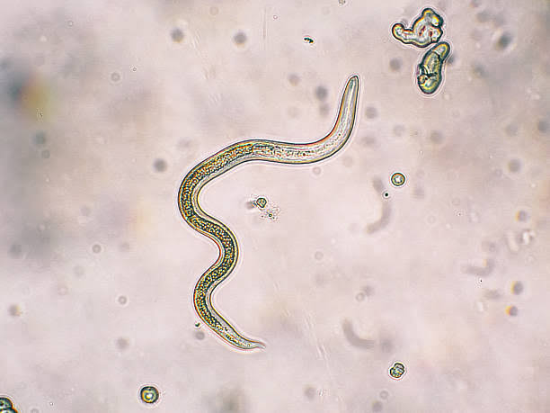
The cave-dwelling olm the bacteria Pelagibacter ubique, which is the most numerous creature in the seas and lichens with their incredibly low metabolism are all examples of oligotrophic species.

Caulobacter crescentus is an oligotrophic Gram-negative bacteria found in freshwater waterbodies. The whole cell functions as an integrated system in the control circuitry that controls and paces Caulobacter cell cycle development. As it orchestrates activation of cell cycle subsystems and Caulobacter crescentus asymmetric cell division, the control circuitry monitors the environment and the internal status of the cell, including the cell topology. The control system has been meticulously tuned as a whole system for reliable functioning in the face of internal stochastic noise and external unpredictability by evolutionary selection.

The bacterial cell’s control system is organised in a hierarchical manner. The signalling and control subsystem communicates with the outside world through sensory units that are mostly found on the cell surface. To adjust the cell to current conditions, the genetic network logic responds to signals received from the environment as well as internal cell status sensors.

ENVIRONMENT AND LOCATIONS

Oligotrophic lakes are often found in northern Minnesota, with deep clear water, stony or sandy bottoms, and minimal algae.

Oxygen levels are high throughout the water column in oligotrophic lakes. Cold water may store more dissolved oxygen than warm water, thus oligotrophic lakes’ deep regions remain quite cold. Low algal content also provides for more light penetration and less breakdown. Algae, zooplankton, and fish die and are degraded by bacteria and invertebrates at the bottom of the ocean. The process of breakdown consumes oxygen. 

Locations

 Oligotrophs and eutrophs coexist in natural ecosystems, and their proportions are determined by an individual’s capacity to prevail in a given environment.  Despite their capacity to exist in low-nutrient settings, they may struggle to survive in nutritionally- rich ones. Most microorganisms are not well adapted to exist in nutrient-limited circumstances and frigid temperatures (below 5 °C), Antarctic habitats offer very little to sustain life. Some of the documented examples of oligotrophic environments in Antarctica are:

Lake Vostok, a freshwater lake cut off from the rest of the world by 4 kilometres (2.5 miles) of Antarctic ice, is often cited as a prime example of an oligotrophic ecosystem. Because of the lake’s severe oligotrophy, some people assume that sections of it are entirely sterile. This may be used as a model to simulate alien life investigations on frozen planets and other celestial worlds.

Oligotrophic soil environments

In general, nutrient availability decreases as the depth of the soil environment increases, since organic molecules degraded from detritus are swiftly eaten by other microorganisms on the surface, resulting in nutritional deficiency in the deeper levels of soil.

Collimonas is one of those species that may survive in an oligotrophic environment as it has the capacity to not only hydrolyze the chitin generated by fungus for nutrition, but also to create materials. Fungi are a prevalent element of the habitats where Collimonas thrives. In oligotrophic settings, reciprocal relationships are prevalent. Weathering also allows Collimonas to access electron sources from rocks and minerals.

The environment of soil in polar locations, such as the Antarctic and Arctic regions, is termed oligotrophic since the soil is frozen and biological activity is minimal. Actinobacteria, Proteobacteria, and Cyanobacteria are the most common bacteria in frozen soil, with a tiny quantity of archaea and fungus. Under a wide range of low temperatures, actinobacteria can keep their metabolic enzymes active and continue their biochemical processes.

The following are the characteristics that a bacterium should have in order to be labelled as an oligotroph:

(a) Having a form with a high surface-to-volume ratio.

(b) Having an innate propensity for using metabolic energy for food absorption during phases of growth stagnation.

(c) Possessing nutrition absorption abilities that are expressed in a constitutive manner.

(d) Presence of a low-specificity, high-affinity transport mechanism that allows for simultaneous absorption of mixed substrate.

 (e) Having systems for conserving nutrition after it has been absorbed.

Extremophiles and their products have revolutionised many aspects of our home and professional life, from household materials to molecular diagnostics. It is not unlikely that new and medically useful discoveries will be found in the realm of extremophile research; the potential of these organisms is so fresh and huge that their applications may be restricted only by imaginations.

SPOILAGE OF MEAT

BY DAKSHITA NAITHANI

INTRODUCTION

Food spoiling is described as any alteration that the customer finds objectionable. Spoilage happens at any given point of time in the food chain. Insect damage, physical damage, indigenous enzyme activity in animal or plant tissue, and microbial infections can all contribute to spoilage. The majority of natural products have a definite shelf life. 

Meat and its derivatives are nutrient-dense foods that are devoured by people all over the world.

Meat deteriorates biologically and chemically from the time it is slaughtered until it is consumed. Microbial spoilage may occur in meat and its products such as ham, sausages, cooked meat, dried meats, minced meat, and so on.

Contamination source and causes

Natural processes such as oxidation or autolytic activity in the muscle following slaughtering can cause meat decomposition. Microbial contamination of meat is caused by a number of variables, including the animal’s microflora, the type of container used, and how the meat is handled and stored. Knives, utensils, hands, and workers’ clothes, among other things

Numbers of microbes of microbes resides on the meat and their products. Some of important are listed below:

 Brochothrix thermosphacta

It can grow in both aerobic and anaerobic environments, and meat is a niche for it. This microorganism is frequently found in irradiated meat and poultry and is responsible for the bad odour of meat.

Carnobacterium

Carnobacterium is a gram-positive bacterium with nine different species. C. divergens has been demonstrated to show green discoloration of ham as a result of H2O2 generation.

Clostridium tetani

Clostridium creates a huge quantity of gas in packed meat, which, when combined with bad smells, causes the container to blow open.

Enterobacteriaceae

These bacteria can act as facultative anaerobes, oxidase-negative glucose fermenters, and nitrate suppressants, and they can influence to the rotting of meat.

Leuconostoc

D-lactate and ethanol are produced by Leuconostoc, a lactic acid generating bacterium. The discolouration, gas generation, and buttery odour of rotting meat are aided by these bacteria.

Pseudomonas

Pseudomonas has been identified as the most common bacterium found in rotting meat. Pseudomonas is widely recognised for successfully exploiting meat as a niche due to its capacity to break down glucose and amino acids under aerobic and cold conditions.

The spoilage of different types of meat are :

Spoilage of fresh meat

The enzymes and microbial activity in fresh meat causes it to decay. Proteolytic effects on muscle and connective tissue, as well as fat hydrolysis, are caused by autolysis alterations. Salmonella, E.coli, Listeria and other microbial diseases have been identified in fresh meat.

Spoilage of meat

Rotting of meat is caused by three fundamental mechanisms: microbial growth, oxidation, and enzymatic autolysis. Meat is a great substrate for microbial development due to its nutritional makeup, high water content, and mild pH. Staphylococcus, Streptococcus, Clostridium, and Salmonella are among the bacteria found in an animal’s lymph nodes, which might influence meat.

Spoilage of refrigerated meat

Fresh meat may be present in good condition for 5-7 days when refrigerated at 4°C. The development of psychrophilic microbes is favoured by a cool environment over time. Contamination of meat by rotting and dangerous bacteria can occur due to poor hygiene. B. thermosphacta and lactic acid bacteria are the microbes that cause refrigerated meat deterioration in general.

Factors spoilage of meat and meat products

Buffer capacity and pH

Muscle pH drops to 5.4-5.8 after slaughtering, however meat from stressed animals and cooked products, such as sliced ham, have pH levels greater than 6. The presence of tissue and a high pH in meat results in a faster deterioration process owing to microbial growth and nutritional absorption.

Water activity

The quantity of water in a meal that is available for the growth of microorganisms, including diseases, is measured by water activity (aw). Raw meat has a value of 0.98-0.99, while cooked meat has a value of 0.94; these values enable most bacteria to thrive. Pathogens cannot develop or produce toxins if the water activity is less than 0.85.

Packaging and gaseous atmosphere

The constitution of rotting flora is heavily influenced by packaging circumstances and the gaseous content of the environment surrounding the meat. Aerobic conditions favour the development of Pseudomonads over everything else. Lactic acid bacteria are the most prevalent microbes in vacuum or CO2-modified environment packed goods, and they are the bacteria that cause the most deterioration.

Storage temperature

The duration of the lag phase, the maximum specific growth rate, and the ultimate cell number are all affected by temperature. Lower temperatures inhibit microbial contamination and alter the microbiota makeup of meat. The dominance of lactic acid bacteria in sealed beef products is likewise maintained in cold circumstances. In sealed refrigerated beef, psychrophilic Clostridium spp. were found. The growth of Enterobacteriaceae, Pseudomonas spp., and Acinetobacter spp. is influenced by abuse of temperature.

General types of Spoilage of meats and meat Products.

Spoilage under Aerobic condition

The spoilage of meats and meat products due bacterial in aerobic conditions are:

 Surface slime: Surface slime generated by Pseudomonas, Acinetobacter, Micrococcus, Streptococcus, Leuconostoc, and Bacillus results in spoilage. Lactobacillus species have also been shown to generate slime. Microbes are benefited from the thin layer development on meat because it delivers nutrients from the substrate.

Changes in colour of meat pigments: As a result of bacteria producing oxidising chemicals such as Peroxides or Hydrogen Sulphide, the red colour of meat, known as its “bloom,” might change to green, brown, or grey. Greening of sausage has been linked to Lactobacillus which is mainly hetero fermentative and Leuconostoc species.

Changes in fats: The oxidation of lipids in meat occurs chemically in the presence of oxygen and can be accelerated by light and copper. Lipolytic bacteria can induce some lipolysis as well as speed up the oxidation of lipids. Fat rancidity can be induced by lipolytic Pseudomonas and Achromobacter species.

Phosphorescence: Luminous bacteria, such as Photobacterium spp., develop on the surface of the flesh and create this rare abnormality. Other bacteria that generate red pigments might be responsible for the red spot.

Off odours and off tastes: Unpleasant odours and taste develop in meat as a result of bacteria growing on the surface. They emerge before other symptoms of deterioration. Meat sourness can be induced by volatile acids such as formic, acetic, butyric, and propionic acids, as well as yeast proliferation.

Spoilage due to moulds in aerobic condition

Moulds are actively involved the spoilage meat in aerobic conditions. Common types of spoilage are listed below.

Stickiness: Stickiness is caused by fungus bud development, which makes the surface of the flesh sticky to the touch. As a result of this form of deterioration, flesh takes on an odd look.

Whiskers: In the absence of sporulation, a little amount of mycelial development can occur resulting in whisker growth. Moulds such as Thamnidium elegans, Mucor mucedo, M. lusitanicus, or M. racemosus, Rhizopus, and others can create this sort of fuzzy growth.

Black spot and White spot: Cladosporium herbarum is the most prevalent source of this sort of spot, although other moulds with dark colours might also be to fault. White spot is most frequently reported by Sporotrichum carnis, although it can also be caused by any mould with moist, yeast-like colonies, such as Geotrichum.

Green patches:  Species of Penicillium such as P. expansum, P. asperulum, and P. oxalicum are responsible for the green patches on meat.

Decomposition of fats:  The oxidation of fats is caused by the hydrolysis of fats caused by lipase produced by moulds.

Spots on surface: Yeast and mould spoilage is generally confined to a large extent and may be cut away without harming the remainder of the meat.

Spoilage under Anaerobic Conditions

Facultative and anaerobic bacteria are able to grow within the meat under anaerobic conditions and cause spoilage. Few are listed below:

Souring: It can be generated by the meat’s natural enzymes during age or ripening, bacterial synthesis of fatty acids or lactic acid, or proteolysis without putrefaction induced by facultative or anaerobic bacteria and frequently referred to as “stinking sour fermentation.” Clostridium species and fecal coliform feed on carbohydrates and produce acid and gas. The development of lactic acid bacteria is prevalent in vacuum sealed meats, especially in gastight wrappers.

 Putrefaction: Putrefaction is the disintegration of protein caused by anaerobic bacteria producing foul-smelling chemicals including hydrogen sulphide, indole, skatole, ammonia, and amines. In most cases, Clostridium spp. are to blame, however facultative bacteria can also cause or contribute to putrefaction. Clostridia-induced putrefaction is frequently accompanied by gas production.

Taint:  Having a foul odour and a poor taste. Temperature, in addition to air, has a significant impact on meat decomposition. Many microbes, including Pseudomonas, Lactobacillus, Leuconostoc, Streptococcus, and Flavobacterium species, generate slimes, discoloration, and growth patches on the surface and can induce sourness.

BACTERIAL MENINGITIS

BY- DAKSHITA NAITHANI

INTRODUCTION

The inflammation of the meninges is known as meningitis. The Dura mater, arachnoid mater, and pia mater are the three membranes (meninges) that border the vertebral canal and skull, encapsulating the brain and spinal cord. Symptoms such as headaches, fever, and stiff neck are common.

Prior to the discovery of antibiotics, this was a fatal illness. Despite tremendous advancements in healthcare, the disease still has a death rate of over 25%. Many different pathogens can cause the disease, but bacterial meningitis has the largest worldwide impact.

Despite advances in diagnosis, treatment, and immunisation, 8.7 million cases of meningitis were recorded globally in 2015, with fatalities as much as 379,000. In early 2020, the first incidence of meningitis linked to COVID 19 was discovered. Every year on April 24th, World Meningitis Day is commemorated. Meningitis is one of the leading causes of illness and death in children under the age of five worldwide. According to Indian studies, meningitis is one of the main causes of mortality among infants under the age of five. 

TYPES OF MENINGITIS

Viral meningitis: It is the most frequent, but not the most dangerous, form of meningitis, accounting for 85 percent of cases. Enteroviruses are among the most prevalent causing viruses.

Bacterial meningitis: Bacterial meningitis is the second most prevalent kind of meningitis, affecting around 3 per million individuals each year. N. meningitidis, S. pneumoniae, H. influenzae, and S. aureus are the microorganisms that cause this kind of meningitis. Inflammation of the meninges can be caused by the same bacterium that causes TB.

In most countries, N.meningitidis is the primary cause of meningitis and a feared illness. The fatality rate from bacterial meningitis is frequently greater in underdeveloped nations than in industrialised countries.

Fungal meningitis: It’s a rare occurrence that generally leads to persistent meningitis. It is caused by a fungus that infects the body and travels from the blood to the nervous system, as the name implies.

Parasitic meningitis: It is less frequent than viral or bacterial meningitis and is caused mostly by parasites found in soil, excrement, cereals, or chickens. The infection is spread through ingesting the parasite’s eggs rather than normal routes. One of the most severe diseases is amoebic meningitis.

Non-infectious meningitis: It is a complication of an underlying health condition, rather than an infection. Inflammation in the tissues can be caused by a variety of factors, including drug use, head trauma, brain surgery, and cancer-related issues.

HOW DOES IT SPREAD FROM ONE INDIVIDUAL TO ANOTHER?

Meningitis caused by fungi, parasites, or non-infectious organisms is not contagious, while viral and bacterial are extremely contagious. Sneezing, coughing, and sharing utensils, cutlery, and toothbrushes are all ways to spread viral and bacterial meningitis. People who have these viruses or bacteria in their nose or throat but are not ill are generally carriers.

RISK FACTORS FOR MENINGITIS:

Risk factors for meningitis include:

•People who do not complete or skip their recommend childhood or adult immunization schedule

• Most of the viral cases occur in children younger than five years of age. Bacterial cases are common to those under the age of twenty years. Age also plays a big role in determining the risk factor.

 • It is possible to live in a community. Meningococcal meningitis is more common in college students who live in dorms and children who attend boarding schools or child care centres. This is most likely due to the bacterium’s ability to spread fast among big populations through the respiratory pathway.            

• Immune system dysfunction. Meningitis is also made more likely by AIDS, alcoholism, diabetes, immunosuppressive medications, and other immune system disorders. Anyone without a spleen should be immunised to reduce their risk.

SYMPTOMS

Meningitis affects more than two-thirds of children under the age of two, with the majority of cases occurring in the first two years of life. This might be related to low immunity and increased brain vascularity, which puts children at a higher risk. Furthermore, due to the immaturity of the central nervous system (CNS) in babies and children, the symptoms of infection are also hazy. Due to these reasons doctors depend more on the diagnostic tests rather than the symptoms.

-Fever for more than a week

-Neck stiffness

-Headaches

-Nausea and vomiting

-Altered or reduced level of consciousness

-Lethargy

-Rash

-Convulsions

Meningitis rash

A mild rash is one of the late indicators that one of the bacteria that causes meningitis, Neisseria meningitidis, is present in your circulation. The rash will become more visible as the illness progresses and spreads. The palms of the hands and the inside of the mouth, for example, may exhibit indications of a rash more easily than other parts of the body.

TREATMENT AND MANAGEMENT OF DISEASE

The therapy is determined on the underlying aetiology of meningitis. Antibiotics are used to treat bacterial meningitis, which may necessitate urgent hospitalisation. This might aid in the prevention of brain injury. The treatment of fungus meningitis may need the use of antifungal medicines. Viral meningitis may go away on its own, but you’ll need to see a doctor to figure out what’s causing it and how to treat it properly. On the basis of symptoms present parasitic meningitis is treated.

In all instances of bacterial meningitis, prompt treatment and supportive care and antibiotics are essential. Antibiotics are chosen depending on the organism that is thought to be causing the illness. In order to give the optimum antimicrobial coverage, the physician must consider the patient’s medical history.

Steroid Therapy: There isn’t enough data to back up the use of them in bacterial meningitis.

Chemoprophylaxis: Close contacts of a patient with N. meningitidis and H. influenzae type B meningitis should take this medication. People who have shared utensils, and health care providers in close proximity to secretions are all examples of close interactions.

Physical Therapy Management

In most cases, physical rehabilitation begins in the intensive care unit. It’s critical to remember a patient’s chart contraindications to therapy, such as intracranial pressure, cerebral pressure, and other lab results that dictate rehabilitation recommendations, while starting a plan of care. In the acute phase, proper posture and range of exercises should be started as soon as it is safe to do so. Proper pillow and towel placement will maintain the integrity of the skin and avoid contractures. Maintaining trunk and neck mobility is critical for functional mobility.

The earlier a patient begins therapy, the lower the risk of subsequent impairments, allowing for a better treatment.

If left untreated it can lead to significant brain problems and is sometimes deadly. In 10–20 percent of survivors, it can cause brain damage, hearing loss, or learning disabilities, as well as amputations in certain cases.

DIAGNOSTIC TESTS

 It is detected by analysing the cerebrospinal fluid, which includes a white blood cell count, glucose, protein, and, in rare circumstances, a polymerase chain reaction (PCR). A lumbar puncture is used to collect CSF, and the opening pressure can be monitored.

Bacteria in the blood are identified using blood cultures. Bacteria have the ability to move from the bloodstream to the meninges. Both sepsis and meningitis can be caused by a variety of bacteria, including N. meningitidis and S. pneumonia.

A differential complete blood count is an indicator of health general. The amount of red and white blood cells in your blood is counted. Infection is fought by white blood cells. In meningitis, the count is generally high.

Pneumonia, TB, and fungal infections can all be detected using chest X-rays. Meningitis can develop as a result of pneumonia.

A head CT scan may reveal issues such as a brain abscess. From the sinuses to the meninges, bacteria can spread.

A glass test may also be performed by doctors. The doctor performs this test by rolling a glass over the rash formed. It’s most probable meningitis rash if the rash doesn’t disappear with pressure. The odd patches on the skin may be the consequence of another ailment if it fades away.

PREVENTION

Microbes that cause meningitis can easily be dispersed all around through aerosols when a carrier coughs, sneezes or shares utensils or other oral items. A few steps which are present to prevent it are:-

• Please wash your hands. Hand cleaning is important in preventing the transmission of germs. Hands should be washed before and after meals, touching objects or animals in public places. Show children how to wash and rinse their hands completely and properly.

 •Practice good oral hygiene. Do not share edibles and utensils with anybody else.

•Maintain a good immune system by keeping a good diet and staying healthy.

 •One should always cover their mouth while coughing or sneeze.

• If you’re expecting a child, be cautious about what you eat. Reduce your risk of listeriosis by cooking meat to 165 degrees Fahrenheit, which includes hot dogs.  Choose pasteurised milk cheeses that are clearly stated on the package.

What are germs?

The term “germ” encompasses an army of tiny terrors, including viruses, fungi, parasites, and bacteria. These “pathogens” all have the ability to spread from victim to victim(called a host). Germs are so small you can see them only through a microscope. They look like spiky blogs, oozing spirals,hairy hotdogs, or other microscopic monsters.

Why are germs bad for us?

These microorganisms hitch a ride into our bodies on the food we eat, in the air we breathe, or through a variety of other methods. Once they have invaded our personal spaces, germs reproduce and create toxic waste, which triggers our body’s most repulsive reactions. They make us sniffle, upchuck, run to the toilet, break out in rashes and fevers, and suffer even more unpleasant symptoms.

How do we get sick from viruses?

Most viruses are frail little things ( unlike bacteria and fungi, viruses are not even alive ) that can multiply only inside a living host ( including animals, plants, and even bacteria). There they spread overwhelming and attacking the host’s immune system and causing all sorts of nasty symptoms. Colds, flus, chicken pox, immune disorders, and measles are caused by viruses. Among the worst is a Ebola, which triggers bleeding and is fatal more than half the people who catch it.

How do we get sick from fungi?

Fungi are microscopic molds, yeasts, and other plant like pathogens that thrive in wet, warm places like our armpits, our belly buttons, and the dank spaces between our toes. They feed on our respect and dead tissues and produce stinky wastes that irritate our skin.

How do we get sick from parasites?

This ghastly germ group includes itty-bitty insect larvae, amoebas, and one celled organisms called Protozoa that live in nasty food, damp soil, or dirty water. Parasites depend on a living host for their survival. They sneak into our bodies in tainted water and food, costing of all sorts of gastrointestinal gripes: diarrhoea, vomiting, upset stomachs, and worse. Malaria – common diseases that causes chills, shaking, and fevers – is spread by a parasite passed in mosquito bites. These life-sucking relationships are often the stuff of nightmares.

How do we get sick from bacteria?

Unlike viruses, bacteria are living single celled organisms that can reproduce both outside and inside the body. Like all living things, bacteria create waste -microscopic poops that can act as a poison inside the host. You can blame sore throat, ear infections and tooth-tartar buildup on bacteria. One of the most famous bacteria is Escherichia coli. This rod shaped micorbe lives deep in your intestines, the body’s busiest bacterial neighborhood. Harmful ones make you puke for days.E.coli strains produce an important vitamin. That’s right – some bacteria are actually good for you!

How many bacteria are inside our body right now?

Your body is built of trillions of itty-bitty living blobs, called cells, that work together to make you you. But for every cell you call your own, ten foreign bacteria cluster around or near it. You are a microbe metropolis! Scientists call these communities of foreign bacteria your body’s “flora”, and no two people host the same mix of microorganisms. In fact, scientists are beginning to think of your flora as just another organ.

Can we see these bacteria?

No, they are microscopic. But you can certainly smell them. Like any living thing, bacteria eat, reproduce,die, and create waste which can make your life stink – literally !(Bacteria are the source of bad breath and body odor.)

Benefits of Bacteria

Your gut reaction might be to wrinkle your nose at the thought of bacteria inside your guts, but it turns out that many so-called good bacteria are essential to your health, the survival of life on Earth, and the making of tasty foods. Behold, the benefits of a microscopic allies…

Health boosting

Your body’s microbes support your immune system, which fights sickness.

Plant feeding

Blue-green algae and other types of bacteria convert the nitrogen in the air into compounds plants can use.

Food processing

Micorbes in our innards play a huge role in the digestive process, helping us absorb nutrients and vitamins from our food.

Food making

Bacteria are a vital ingredient in the process of turning milk into yogurt and tasty cheeses. The holes in Swiss cheese are created by carbon dioxide bubbles exhaled by bacteria during the cheese making process.

Planet Cleaning

Bacteria breakdown dead animals and plants, which “decompose” into nutrients for the living.

References :

WHY?-Answers to everything, Image publications.

ANTI-CELL WALL ANTIBACTERIAL DRUGS

Selective toxicity is the important characteristic of antimicrobial drugs which means that any drug is selective against a particular microorganism and also selectively act on a particular site. Not all drugs can act on every site. There are many sites at which any drug acts such as cell wall, cell membrane of the bacterial cell. Basically selective toxicity explains that any drug will only act on the pathogen and not on the host.
ANTI-CELL WALL DRUGS
Anti-cell drugs are those drugs which act on the cell wall of the bacterial pathogen and not the host. There are variety of drugs which fall under this category. The major class is of beta-lactam antibiotics among which penicillin is the drug which is studied the most. The drugs can be administered into the patient’s body by different ways like intramuscular, intravenous, or can be applied as topical preparations. But mostly, these drugs are intramuscular or intravenous drugs. The following points explain the further different mechanisms of anti-cell wall drugs.
There are 3 different mechanisms by which anti-cell wall drugs work and thus they are also classified as following:

  1. First classification involves the drugs that directly interact with Penicillin-Binding-Proteins (PBPs) and inhibit the transpeptidase activity which in turn inhibits the attachment of newly formed peptidoglycan subunit to the pre-existing one.
    This is the main mechanism of β-lactam antibiotics. These antibiotics include Penicillin (penams), cephalosporins, Penems, Carbapenems, and monobactams.
    These antibiotics bind to the penicillin-binding proteins which are enzymes present in the bacterial cell wall. Different β-lactam antibiotics bind in a different way. After the antibiotics bind to the enzyme, it changes the morphological response of the bacteria to the antibiotic.
  2. Second classification involves the drugs that bind to the peptidoglycan subunit, blocking different processes.
    The important class of compounds called as glycopeptides are mainly involved in this mechanism of anti-cell wall antibiotics.
    Vancomycin and Teicoplanin are the major examples of glycopeptide antibiotics.
    Vancomycin kills only gram-poitive bacteria whereas Teicoplanin is active against both. The overall mode of action of glycopeptides antibiotics is blocking transpeptidation i.e. similar to β-lactam antibiotics, they also inhibit the transpeptidase activity, and transglycosylation i.e. they being large in size attach to the peptidoglycan subunits thus creating a blockage which does not allow the cell wall subunits to attach to the growing peptidoglycan backbone.
  3. Third classification involves the drugs that block the transport of peptidoglycan subunits across cytoplasmic membrane.
    The main example of such type of drugs is bacitracin, which is a simple peptide antibiotic originally isolated from Bacillus subtilis.
    The mode of action of these class of drugs is blocking the activity of specific cell membrane lipid carriers which act as the attachment surface for peptidoglycan precursors and help in their movement from cell cytoplasm to exterior of the cell. This activity of lipid carriers is inhibited by bacitracin like drugs and they finally prevent the incoroporation of those precursors into cell wall thus inhibiting its biosynthesis.

Although, its route of administration is mostly oral or intramuscular, bacitracin is also known to show its effects when used as topical ointments like Neosporin.

GRAM STAINING

Bacteria can be classified into two different categories i.e. gram-positive bacteria and gram-negative bacteria. To differentiate the type of bacteria present in any collected sample there is a technique known as GRAM STAINING OR GRAM STAIN. It is sometimes also known as gram’s method. This technique is a different step process which can easily distinguish and classify between different types of bacteria. This was named after the great scientist Hans Christian Gram.
Gram Staining method differentiates bacteria on the basis of their physical and chemical structure of cell wall. They are stained with different reagents and are observed in different colors when seen under the microscope. It is due to the fact that gram positive bacteria have a thick layer of peptidoglycan which allows it to retain the primary stain which is crystal violet and thus they appear purple whereas on the other hand, gram-positive bacteria have thin peptidoglycan cell wall and thus only retain the secondary or counter stain which is Safranin and thus they appear slightly pinkish when observed under microscope. Gram staining is the basic technique which is widely used in the microbiology labs to distinguish between both the types of bacteria. It provides a great help to microbiologists to perform their clinical tasks. If any infection is suspected in the patient then after the collection of sample, the gram staining is done and the type of bacteria is observed.
Gram staining is completed in the various steps –

  1. Fixation of clinical materials i.e. the sample collected are being fixed on the glass slide by first making a smear using water and then that smear is either heat fixed or methanol fixed. It is a first and a very important step. Methanol fixation was later discovered keeping in mind its advantage of not destroying the morphology of host cell, as well as bacteria present in that. It is majorly used for the testing of blood samples collected from patients.
  2. Application of primary stain i.e. crystal violet. Primary stain means the first stain which is applied onto the fixed smear which stains all the calls purple or blue.
  3. To distinguish the slide is then washed off in a gentle and indirect stream of water for 2 seconds which removes the extra stain that is not absorbed by the cells.
  4. The next step involves the application of a mordant i.e. Iodine solution. This forms a complex with crystal violet due to which all the cells start appearing blue.
  5. Again the slide is washed for 2 seconds to remove extra stain.
  6. Addition of a decolorizing agent is the next step which will ultimately remove the excess stain which bacteria has not absorbed. The decolorizer contains the combination of acetone and alcohol. In this step, the gram positive bacteria continue appearing violet or blue in color whereas gram negative bacteria start appearing colorless.
  7. Application of counter stain or secondary stain i.e. safranin is the next step. It should let remain on the slide for 30 second to 1 minute. This will stain all the colorless gram negative bacteria pink and gram positive bacteria remains blue in color.
  8. Again the slide is washed off in a gentle stream of water.
  9. The prepared stained slide is then observed under a microscope using immersion oil (for observing under 100x).

MECHANISM OF DIFFERENT TYPES OF ANTIBIOTICS

Antibacterial Drugs are classified according to their site of action which are as follows :

CELL WALL SYNTHESIS INHIBITORS
There are 3 different mechanisms by which anti-cell wall drugs work and thus they are also classified as following:

  1. First classification involves the drugs that directly interact with Penicillin-Binding-Proteins (PBPs) and inhibit the transpeptidase activity which in turn inhibits the attachment of newly formed peptidoglycan subunit to the pre-existing one.
    This is the main mechanism of β-lactam antibiotics. These antibiotics include Penicillin (penams), cephalosporins, Penems, Carbapenems, and monobactams.
    These antibiotics bind to the penicillin-binding proteins which are enzymes present in the bacterial cell wall. Different β-lactam antibiotics bind in a different way. After the antibiotics bind to the enzyme, it changes the morphological response of the bacteria to the antibiotic.
  2. Second classification involves the drugs that bind to the peptidoglycan subunit, blocking different processes.
    The important class of compounds called as glycopeptides are mainly involved in this mechanism of anti-cell wall antibiotics.
    Vancomycin and Teicoplanin are the major examples of glycopeptide antibiotics.
    Vancomycin kills only gram-poitive bacteria whereas Teicoplanin is active against both. The overall mode of action of glycopeptides antibiotics is blocking transpeptidation i.e. similar to β-lactam antibiotics, they also inhibit the transpeptidase activity, and transglycosylation i.e. they being large in size attach to the peptidoglycan subunits thus creating a blockage which does not allow the cell wall subunits to attach to the growing peptidoglycan backbone.
  3. Third classification involves the drugs that block the transport of peptidoglycan subunits across cytoplasmic membrane.
    The main example of such type of drugs is bacitracin, which is a simple peptide antibiotic originally isolated from Bacillus subtilis.
    The mode of action of these class of drugs is blocking the activity of specific cell membrane lipid carriers which act as the attachment surface for peptidoglycan precursors and help in their movement from cell cytoplasm to exterior of the cell. This activity of lipid carriers is inhibited by bacitracin like drugs and they finally prevent the incoroporation of those precursors into cell wall thus inhibiting its biosynthesis.

Although, its route of administration is mostly oral or intramuscular, bacitracin is also known to show its effects when used as topical ointments like Neosporin.

INHIBITORS OF PROTEIN SYNTHESIS
Protein Inhibitors can be divided into 2 parts:

  1. Inhibitors binding to 30S subunits
    • Aminoglycosides bind to the bacterial ribosome, after which they cause tRNA mismatching and thus protein mistranslation.
    This occurs by mismatching between codons and anticodons, which synthesize proteins with incorrect amino acid. This mistranslated protein, along with correctly translated proteins move into move into the periplasm where most of the mistranslated proteins are degraded and some of them are inserted into cytoplasmic membrane. This causes disruption of the membrane, ultimately killing the bacterial cells.
    • Tetracyclines are bacteriostatic and block the binding of tRNAs with the ribosome during translation thus inhibiting protein synthesis. Most of the tetracycline class of drugs are broad spectrum and are active against wide range of bacteria.
  2. Inhibitors binding to the 50S subunit
    • Macrolides are the large class of naturally produced secondary antibiotics. They are basically broad spectrum, bacteriostatic antibiotics. Their main mode of action is blocking peptide chain elongation and they inhibit the formation of peptide bond.
    Patients allergic to penicillins are recommended erythromycin which is a macrolide.
    • Lincosamides include lincomycin and clindamycin. Though they are structurally different but functionally similar to macrolides. They are specifically known to inhibit streptococcal and staphylococcal infections.
    • Chloramphenicol also inhibits peptidyl transferase reaction inhibiting peptide bond formation. It was the first broad spectrum antibiotic and is very much active against a broad range of bacterial pathogens but is very toxic and can cause side.

INHIBITORS OF MEMBRANE FUNCTION
Biological cytoplasmic membranes are basically composed of lipids, proteins and lipoproteins. The cytoplasmic membrane acts as a selective barrier which allows the transport of materials between inside the cell and the environment.
A number of antibacterial agents work by targeting the bacterial cell membrane. They basically are involved in the disorganization of the membrane. Polymyxins and Lipopeptides are the main anti- cell membrane agents.

NUCLEIC ACID SYNTHESIS INHIBITORS
These drugs inhibit nucleic acid synthesis function by either of the following:

  1. Interfere with RNA of bacterial cell
    Antibacterial drugs of this mechanism are selective against bacterial pathogenic cells.
    For example: The drug rifampin, belonging to the drug class rifamycin blocks the bacterial RNA polymerase activity. It is also active against Mycobacterium tuberculosis and thus id used in the treatment of tuberculosis infection. It also shows side effects.
  2. Interfere with DNA of bacterial cell
    There are some antibacterial agents that interfere with the activity of DNA gyrase.
    The drug class fluoroquinolones show this mechanism. They are borad spectrum antibacterial agents. Some examples of drugs in fluoroquinolone family are Ciprofloxacin, Ofloxacin, Moxifloxacin, etc

INHIBITORS OF METABOLIC PATHWAYS
There are some antibacterial drugs which act as ANTIMETABOLITES and inhibits the metabolic pathways of bacteria.
• The sulfonamides block the production of dihydrofolic acid.
This blocks the production of purines and pyrimidines required for nucleic acid synthesis by blocking the biosynthesis of folic acid. Their mechanism of action is bacteriostatic and they are broad spectrum antibacterial agents. Though humans also obtain folic acid but these drugs are selective against bacteria.
Sulfones are also structurally and functionally similar to sulfonamides.
• Trimethoprim is used in the same folic acid synthesis pathway but at a different phase, in the production of tetrahydrofolic acid.
• There is another drug, Isoniazid which is an antimetabolite only selective against mycobacteria. It can also be used to treat tuberculosis when used in combination with rifampin and streptomycin.

INHIBITORS OF ATP SYNTHASE
There is a class of drug compounds called as Diarylquinolones that are specifically active against mycobacterial growth. They block the oxidative phosphorylation process and finally leading to reduced ATP production which either kill or inhibit the growth of mycobacterial species.

BASICS OF A MICROBIOLOGY LAB

Microbiology is the study of microbes i.e. the organisms which we can’t see with the naked eyes. Although many microorganisms are beneficial for the human use, some are pathogenic also which causes diseases. Clinical Microbiological Laboratory is concerned with finding of those infectious, pathogenic microbes.

MATERIALS USED IN MICROBIOLOGY LAB
Laminar flow hood, Incubator, Autoclave, Refrigerator, Bunsen Burner, Wire loop, Petri plates, Glass slides, Weighing balance, Media plates, Sensitivity disks, Staining rack, Microscope, Bio safety Cabinet, Centrifuge etc.

INTRODUCTION TO DIFFERENT MEDIA
Some of the media used in the microbiology lab are :

  1. MacCONKEY AGAR : It is the selective and differential media used for the isolation of Gram-negative Bacteria. This media can be used for differentiating Lactose fermenting and Non-lactose fermenting bacteria.
  2. BLOOD AGAR : It is the enriched media for the growth of bacteria such as streptococci.
  3. CHOCOLATE AGAR : It is the lysed Blood Agar. The only difference in blood agar and chocolate agar is that in blood agar RBCs are lysed. This enriched media is suitable for the growth of bacteria that are unable to grow on Blood Agar.
  4. THIOSULFATE CITRATE BILE SALT AGAR (TCBS) : It is the selective as well as differential media for the growth of vibrio cholerae , a causative organism for cholera.

GRAM STAINING
Gram staining is the process for differentiating Gram positive and Gram negative bacteria. When the whole procedure of gram stain is followed and the slide is observed under the microscope, Gram positive bacteria appear Violet in color and Gram negative bacteria appear Pink in color.
For the gram staining we need Glass slide, Normal Saline, Inoculating loop, Bunsen burner, Crystal Violet, Gram’s Iodine, Acetone, Safranine.

PROCESS :

  1. The isolated colony of the microorganism is taken and in the drop of normal saline on the glass slide the colony is mixed with the help of inoculating loop to make a smear. The prepared smear is heat fixed.
  2. A staining rack is taken and on the smear, Crystal Violet is added. After 1 minute, the stain was removed by washing the slide in running water.
  3. After that, Gram’s Iodine is added on the smear as a decolorizing agent which is again washed after 1 minute under the running tap water.
  4. The next step is to add Acetone on the smear which is added in the hand to hand process.
  5. After the decolorisation is done, Safranine is added on the smear which is also washed after 1 minute.
  6. The glass slide is then air dried and observed under the microscope.

RESULTS :
It was observed under the microscope that the Gram positive bacteria appear Violet in color due to Crystal Violet stain whereas Gram negative bacteria appear Pink in color due to safranine.

TESTS ANALYZED
The tests analyzed in the microbiology section of the laboratory are basically the culture and sensitivities tests of urine, stool, sputum, pus swab etc.
The basic procedure of performing all the tests are :

  1. First of all, all the tests are performed inside the laminar flow hood.
  2. The samples collected from the patients and the media plates are kept inside the laminar flow.
  3. The inoculating wire loop is heat sterilized and with the help of it, the samples are cultured or streaked on the media plates.
  4. After inoculation, the cultured media plates are incubated for 24 hours (48 hours if necessary) for allowing the growth of bacteria.
  5. After the growth, staining is done or sensitivities are checked according to the requirement by the doctor.
  6. The report is prepared for the patient.