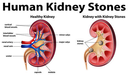
THE EXCRETORY SYSTEM

Excretion

Removal of nirogenous waste produce during metabolism of protein and nucleic acid.

Human excretory system includes:-
Pair of kidney
Pair of ureter
Urinary bladder
Urethra

Function of kidney

Kidneys regulate the osmotic pressure of a mammal’s blood through extensive filtration and purification, in a process known as Osmoregulation.

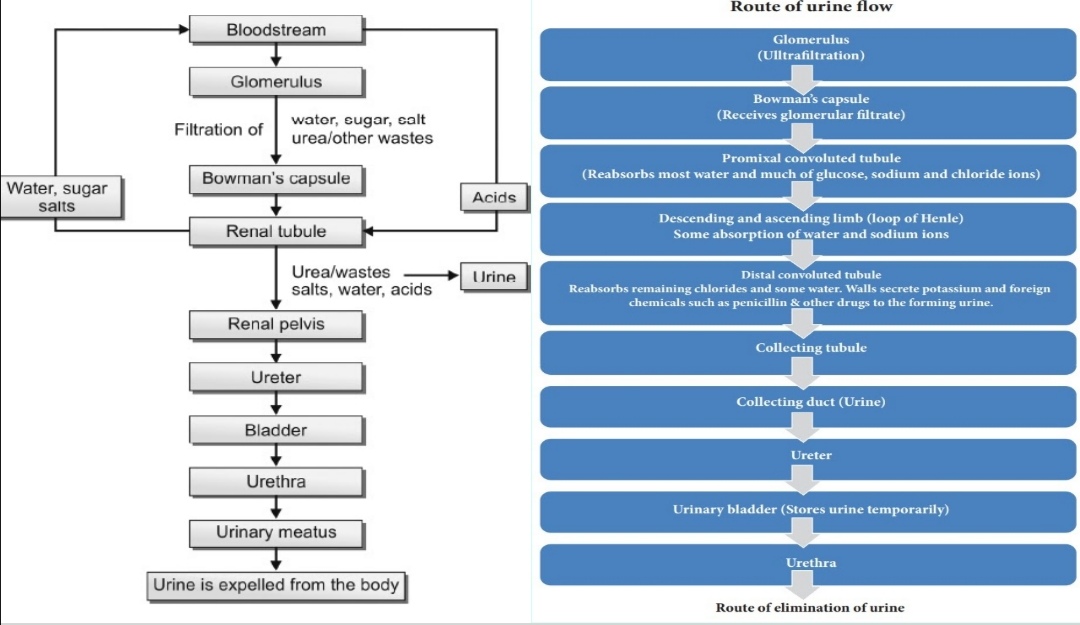
  • Kidneys filter the blood; urine is the filtrate that eliminates wastes from the body via the ureter into the urinary bladder.
  • The kidneys are surrounded by three layers:
    1.  Renal fascia
    2. perirenal fat capsule
    3. Renal caps

EXTERNAL ANATOMY

A typical adult kidney (mass – 135–150 g) is:

10–12 cm- long
5–7 cm – wide
3 cm cm- thick

  • The concave medial border of each kidney faces the vertebral column. 
  • Near the centre of the concave border is an indentation called the Renal hilum, through which the ureter emerges from the kidney along with blood vessels, lymphatic vessels and nerves.
  • Human kidney are Retroperitoneal(covered with peritoneum) 
  • Present between 12th thoracic vertebrae to 3rd lumber vertebrae. 
  • Left kidney is higher than the right kidney due to position of liver in right side. 

Three layers of tissue

a) .The Renal capsule(Deep layer) – Smooth, transparent sheet of dense irregular connective tissue that is continuous with the outer coat of the ureter.

  •  It serves as a barrier against trauma and helps maintain the shape of the kidney. 

b) The adipose capsule (middle layer) – Mass of fatty tissue surrounding the renal capsule. 

  •  Protects the kidney from trauma and holds it firmly in place within the abdominal cavity.

c) The renal fascia(superficial layer) – Thin layer of dense irregular connective tissue 

  •  Anchors the kidney to the surrounding structures and to the abdominal wall 

On the anterior surface of the kidneys, the renal fascia is deep to the peritoneum
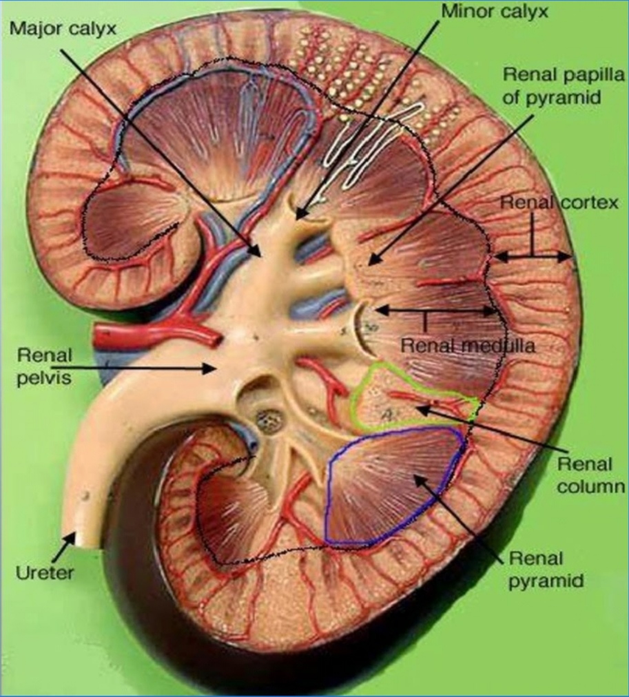
Internal anatomy

A frontal section through the kidney reveals two distinct regions: 

  1.  Renal cortex (outer)
  2.  Renal medulla (inner). 

The renal medulla consists of several cone-shaped renal pyramids. 

The base (wider end) of each pyramid faces the renal cortex, and its apex (narrower end), called a renal papilla, points toward the renal hilum. 

The renal cortex, smooth textured area extending from the renal capsule to the bases of the renal pyramids. 

It is divided into an outer cortical zone and an inner juxtamedullary zone.

Ureter

Muscular tubes of 25-30cm length, 3m in diameter. 

Wall of Ureter

  • Innermost-Transitional epithelium
  • Middle layer-Muscular(longitudinal and circular muscle) 
  • Outermost layer – Tunica adventita. 

Urine is move through ureter by peristalsis.

Urethra conduct the urine from urinary bladder to outside.
Female urethra is short.
Male urethra is long.


Urinary bladder

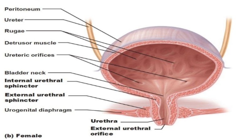
It is hollow muscular organ  that stores urine from the kidneys before disposal by urination. 

In humans the bladder is a hollow distensible organ that sits on the pelvic floor
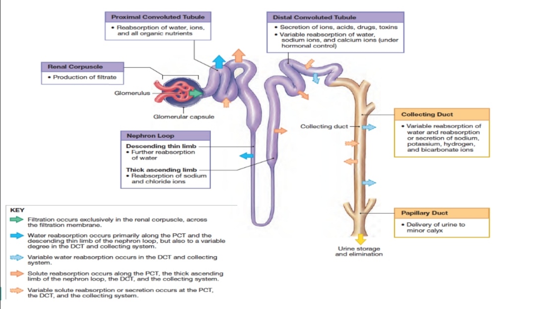
Nephron

Structure and functional unit of kidey.

Each kidney contain about 1 million nephron 

Each nephron has 2 part 

  • Glomerulus
  • Renal tubules.
  • Take a simple filtrate of the blood and modify it into urine.
  • Cleanse the blood and balance the constituents of the circulation.
  • Many changes take place in the different parts of the nephron before urine is created for disposal. 
  • The term urine will be used here after to describe the filtrate as it is modified into true urine. 
  • The principle task of the nephron population is to balance the plasma to homeostatic set points and excrete potential toxins in the urine.

RENAL CORPUSCLE

It CONSISTS OF A GLOMERULUS SURROUNDED BY A BOWMAN’S CAPSULE. 

THE GLOMERULUS ARISES FROM AN AFFERENT ARTERIOLE AND EMPTIES INTO AN EFFERENT ARTERIOLE. 

THE SMALLER DIAMETER OF AN EFFERENT ARTERIOLE HELPS TO MAINTAIN HIGH BLOOD PRESSURE IN THE GLOMERULUS.

THE BOWMAN’S CAPSULE IS DIVIDED INTO THREE LAYERS:

  • OUTER PARIETAL LAYER- MADE UP OF EPITHELIAL CELLS WITH MINUTE PORES OF DIAMETER 12NM.
  • MIDDLE BASEMENT MEMBRANE-IT IS SELECTIVELY PERMEABLE.
  • INNER VISCERAL LAYER-IT CONSISTS OF LARGE NUCLEATED CELLS CALLED PODOCYTE(BEAR FINGER-LIKE PROJECTIONS CALLED PODOCEL) 

Renal tubule

It IS A LONG AND CONVOLUTED STRUCTURE THAT EMERGES FROM THE GLOMERULUS 

IT CAN BE DIVIDED INTO THREE PARTS BASED ON FUNCTION:-

  1.  PROXIMAL CONVOLUTED TUBULE (PCT) –  IN THE RENAL CORTEX.
  2.  THE LOOP OF HENLE, OR NEPHRITIC LOOP – IT FORMS A LOOP (WITH DESCENDING AND ASCENDING LIMBS) THAT GOES THROUGH THE RENAL  MEDULLA.
  3. DISTAL CONVOLUTED TUBULE (DCT)- IN THE RENAL CORTEX.

Loop of Henle

Thick segment-Simple cuboidal epithelium
Thin segment-Simple squamous epithelium.

DCT

Distal  convoluted tubules

Cuboidal epithelium with fewer mitochondria and microvilli

  • Conditional reabsorption of water under the effect of ADH.
  • Na+ – Aldesteron
  • Ca²+ – parathyroid hormone
  • Reabsorption ofHCO3¯and secretion of H+, K+and NH3 to maintain pH. 

Collecting duct

Cuboidal epithelium

Conditional reabsorption of water, Na+, Ca²+. 

Permeability for urea 

PCT

⅔rd of water reabsorption and 60% of glomerular filtrate is reabsorbed. 

Water, Na+, Cl-, HCO3-, Glucose, vit. C, amino acid, K+and little amount of urea and uric acid.

Descending Limb-permeable to water only. 

Ascending Limb– permeable to ions only. 

Na+, Cl-, k+, Mg²+, Ca²+

Reabsorption is minimum. 
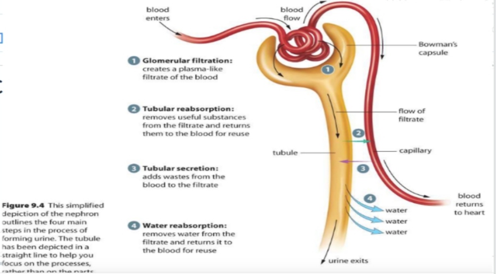
Urine formation

1) Glomerular filtration
2) Tubular reabsorption
3) Tubular secretion

Glomerular filtration 

  • WATER AND SOLUTES ARE FORCED THROUGH THE CAPILLARY WALLS OF THE Glomerulus INTO THE BOWMAN’S CAPSULE (GLOMERULARCAPSULE)
  • FILTRATE –THE FLUID THAT IS FILTERED OUT INTO BOWMAN’S CAPSULE. 
  • Glomerulus filtrate-same as plasma but protein are absent. 

Contains-Water, ions, Glucose, amino acid, water soluble vitamin, urea, uric acid etc. 

Tubular reabsorption

OCCURS BOTH PASSIVE AND Actively. 

GLUCOSE, AMINO ACIDS, AND OTHER NEEDED IONS (NA, K, CL, CA, HCO3) ARE TRANSPORTED OUT OF THE FILTRATE INTO THE PERITUBULAR CAPILLARIES ( REABSORBED BACK INTO THE BLOOD)

ABOUT 65% OF THE FILTRATE IS REABSORBED IN THE PCT. 

AS THESE SUBSTANCES ARE Reabsorbed, THE BLOOD BECOMES HYPERTONICSO WATER EASILY FOLLOWS BY OSMOSIS. 

REABSORPTIONIN THE DCT IS UNDER Hormonal CONTROL ALDOSTERONE CAUSES MORE SALT TO BE ABSORBED

 ADH CAUSES MORE WATER TO BE ABSORBED

TUBULAR SECRETION

WASTE PRODUCTS SUCH AS UREA AND URIC ACID, DRUGS AND HYDROGEN AND BICARBONATE IONS ARE MOVE OUT OF THE PERITUBULARCAPILLARIES INTO THE FILTRATE; THIS REMOVES UNWANTED WASTES AND HELPS REGULATE PH

Urine

It is pale yellow in colour due to Urochrome pigment that is byproduct of red blood corpuscles(RBC) breakdown. 

Around 1-1.5 litre of urine is formed per day. 

PH =6 (vary 4.2 – 8.2) 

It can be four times as concentrated as the blood i. e-1200mosmol/L.

Heavier than water

  • contain
  • 95% =water
  •  5%= urea, uric acid, K+, H+, NH4+, sulphate, hippuric acid, oxalate


Genetic Disorders

Pedigree Analysis

The record of occurrence of a trait in several generations of a human family is called pedigree analysis. The inheritance of a particular trait is represented in the family tree over generations.

Mendelian Disorders

Mendelian disorders are mainly determined by alteration or mutation in the single gene. Mendelian disorders can be traced in a family by the pedigree analysis.

Most common and prevalent Mendelian disorders are :

  • Haemophilia
  • Cystic fibrosis
  • Sickle-cell anaemia
  • Colour blindness
  • Phenylketonuria
  • Thalesemia

Haemophilia

This sex linked recessive disease, which shows its transmission from unaffected carrier female to some of the male progeny has been widely studied. In this disease, a single protein that is a part of the cascade of proteins involved in the clotting of blood is affected. Due to this, in an affected individual a simple cut will result in non- stop bleeding.

The heterozygous female (carrier) for haemophilia may transmit the disease to sons. The possibility of a female becoming a haemophilic is extremely rare because mother of such a female has to be at last carrier and the father should be haemophilic.

Sickle-cell anaemia

This is an autosome linked recessive trait that can be transmitted from parents to the offspring when both the partners are carrier for the gene(or heterozygous). Heterozygous individuals appear apparently unaffected but they are carrier of the disease as there is 50 per cent probability of transmission of the mutant gene to the progeny, thus exhibiting sickle-cell trait.

The defect is caused by the substitution of Glutamic acid by Valine at the sixth position of the beta globin chain of the haemoglobin molecule.

Phenylketonuria

This inborn error of metabolism is also inherited as the autosomal recessive trait.the affected individual lacks an enzyme that converts the amino acid phenylalanine into tyrosine. As a result of this phenylalanine is accumulated and converted into phenylpyruvic acid and other derivatives.

Accumulation of these in brain results in mental retardation. These are also excreted through urine because of its poor absorption of kidney.

Chromosomal Disorders

The chromosomal disorders on the other hand are caused due to absence or excess or abnormal arrangement of one or more chromosomes. Failure of segregation of chromatids during cell division cycle results in the rain or loss of a chromosome, called Aneuploidy. Failure of cytokinesis after telophase stage of cell division results in an increase in a whole set of chromosomes in an organism and this phenomenon is known as Polyploidy.

Down’s Syndrome

The cause of this genetic disorders is the presence of an additional copy of the chromosome number 21.

The affected individual is short statured with small round head, furrowed tongue and partially open mouth. Palm is broad with characteristic palm crease. Physical, psychomotor and mental development is retarded.

Klinefelter’s Syndrome

This genetic disorder is also caused due to the presence of an additional copy of X-chromosome resulting into a karyotype of 47, XXY. Such an individual has overall masculine development, however, the feminine development (development of breast) is also expressed. Such individuals are sterile.

Turner’s Syndrome

Such a disorder is caused due to the absence of one of the X chromosomes, i.e., 45 with X0. Such females are sterile as ovaries are rudimentary besides other features including lack of other secondary sexual characters.

Human Physiology

Human physiology is the study of how the human body functions. This includes the mechanical, physical, bioelectrical, and biochemical functions of humans in good health, from organs to the cells of which they are composed. It serves as the foundation of modern medicine. The human body consists of many interacting systems of organs. These interact to maintain homeostasis, keeping the body in a stable state with safe levels of substances such as sugar and oxygen in the blood. Each system contributes to homeostasis, of itself, other systems, and the entire body. Some combined systems are referred to by joint names. For example, the nervous system and the endocrine system operate together as the neuroendocrine system

As a discipline, it connects science, medicine, and health, and creates a framework for understanding how the human body adapts to stresses, physical activity, and disease. Human physiology is closely related to anatomy, in that anatomy is the study of form, physiology is the study of function, and there is an intrinsic link between form and function. The study of human physiology integrates knowledge across many levels, including biochemistry, cell physiology, organ systems, and the body as a whole.

History of Human Physiology

The study of human physiology began with Hippocrates in Ancient Greece, around 420 BCE, and with Aristotle (384–322 BCE) who applied critical thinking and emphasis on the relationship between structure and function. Galen (ca. 126–199) was the first to use experiments to probe the body’s functions. The term physiology was introduced by the French physician Jean Fernel (1497–1558).In the 17th century, William Harvey (1578–1657) described the circulatory system, pioneering the combination of close observation with careful experiment. In the 19th century, physiological knowledge began to accumulate at a rapid rate with the cell theory of Matthias Schleiden and Theodor Schwann in 1838, that organisms are made up of cells. Claude Bernard (1813–1878) created the concept of the milieu interieur (internal environment), which Walter Cannon (1871–1945) later said was regulated to a steady state in homeostasis.

Study of human physiology

The major systems covered in the study of human physiology are as follows:

  • Circulatory system – including the heart, the blood vessels, properties of the blood, and how circulation works in sickness and health.
  • Digestive/excretory system – charting the movement of solids from the mouth to the anus; this includes study of the spleen, liver, and pancreas, the conversion of food into fuel and its final exit from the body.
  • Endocrine system – the study of endocrine hormones that carry signals throughout the organism, helping it to respond in concert. The principal endocrine glands – the pituitary, thyroid, adrenals, pancreas, parathyroids, and gonads – are a major focus, but nearly all organs release endocrine hormones.
  • Immune system – the body’s natural defense system is comprised of white blood cells, the thymus, and lymph systems. A complex array of receptors and molecules combine to protect the host from attacks by pathogens. Molecules such as antibodies and cytokines feature heavily.
  • Integumentary system – the skin, hair, nails, sweat glands, and sebaceous glands (secreting an oily or waxy substance).
  • Musculoskeletal system – the skeleton and muscles, tendons, ligaments, and cartilage. Bone marrow – where red blood cells are made – and how bones store calcium and phosphate are included.
  • Nervous system – the central nervous system (brain and spinal cord) and the peripheral nervous system. Study of the nervous system includes research into the senses, memory, emotion, movement, and thought.
  • Renal/urinary system – including the kidneys, ureters, bladder, and urethra, this system removes water from the blood, produces urine, and carries away waste.
  • Reproductive system – consisting of the gonads and the sex organs. Study of this system also includes investigating the way a fetus is created and nurtured for 9 months.
  • Respiratory system – consisting of the nose, nasopharynx, trachea, and lungs. This system brings in oxygen and expels carbon dioxide and water.