ASPERGILLOSIS

BY DAKSHITA NAITHANI

INTRODUCTION

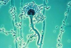
Pier Antonio Micheli, a priest and scientist, named Aspergillus while documenting moulds in 1729. The mould looked like an aspergillum, which is a type of holy water sprinkler. Physician Georg W. Fresenius described the species fumigatus for the first time in 1863.

 Apergillus spp., found in abundance of nature, can be found in soil, decomposing materials, water sources, and air particle. Aspergillus-related disease is uncommon, although it has a significant death rate. The majority of people are exposed to spores (conidia) during their lives, although illness development occurs mostly in people with compromised immune systems, as well as during construction and yard activities. In patients with leukaemia, asthma, cystic fibrosis, and hematopoietic stem cell transplant recipients, A.fumigatus is the most frequent invasive mould infection, with death rates exceeding 50 percent in these high-risk populations.

Around the apex, the stipes are grey in colour. They feature a slick finish. Their globule is tiny and columnar. The conidia’s texture is either smooth or spinose.

WHERE IT CAN BE FOUND?

Aspergillus is commonly found in rotting plant matter. After considerable environmental exposure to Aspergillus spores, such as when handling tree-bark chipping, Aspergillosis can develop in healthy hosts.

Aspergillus can be found in soil, compost piles, and damp grain in outdoor settings. Aspergillus may be found within buildings in wet insulation, fireproofing material, beds , behind couches, in damp rooms’ corners, dust, and air conditioning systems. In the Northern Hemisphere, most research studying seasonal changes in fungal exposure show an increase in airborne Aspergillus throughout the cold season.

TYPES OF ASPERGILLOSIS

Allergic bronchopulmonary aspergillosis (ABPA) is a multifaceted hypersensitivity reaction to inhaled Aspergillus fumigatus. Although it produces lung irritation and allergy symptoms, it does not cause infection. Aspergillus sinusitis is caused by an allergic reaction to the fungus Aspergillus, which produces inflammation in the sinuses and the symptoms of a sinus infection but does not cause an infection. A fungus ball is also known as an Aspergilloma. It forms in the lungs or sinuses but seldom spreads to other regions of the body, as the name implies.

Aspergillosis of the lungs (chronic pulmonary aspergillosis) Aspergilloma, chronic cavitary pulmonary aspergillosis (CCPA), chronic fibrosing pulmonary aspergillosis (CFPA), and Aspergillus nodules are all illnesses classified as chronic pulmonary aspergillosis (CPA). CPA is a lung infection that develops over time.

INVASIVE PULMONARY ASPERGILLOSIS

Inhaled conidia are removed by epithelial cells and alveolar macrophages in normal host lungs. Aspergillus becomes invasive when conidia escape these host defences and develop into branching filaments called hyphae. Alveolar macrophages generate inflammatory mediators, which attract neutrophils, which can destroy the hyphae.

CUTANEOUS SKIN ASPERGILLOS

Aspergillus comes in contact through a breach in the skin (for example, after surgery or a burn wound) and infects persons with weaker immune systems. Cutaneous Aspergillosis can also develop if invasive Aspergillosis moves from another part of the body, such as the lungs, to the skin.

LIFE CYCLE

Aspergillus begins its infectious life cycle by producing conidia (asexual spores) that are easily transported into the atmosphere, ensuring its ubiquity in both habitats. Inhalation of these conidia is the most common way to become infected, followed by conidial deposition in the bronchiole spaces. The phagocytosis and destruction of Aspergillus conidia is largely carried out by macrophages. The danger of infection stems largely from a breakdown in these host defences, as well as fungal characteristics that allow A. fumigatus to survive and flourish in this pulmonary habitat.

SYMPTOMS

The symptoms of Aspergillosis are:

  • Wheezing, chest pain
  • Shortness of breath, stuffiness, runny nose and reduced ability to smell
  • Cough (sometimes blood)
  • Fever (in rare cases), headache, fatigue
  •        Weight loss

•             The length of time that an illness lasts depends on the underlying health concerns, the intensity or location of infection, and the kind of sickness.

•             Allergic forms, may require steroid and antifungal treatment for a few months, possibly longer sometimes.       

•             Invasive pulmonary Aspergillosis may need at least 6 to 12 weeks of antifungal therapy. Severe cases may need therapy for lifetime and monitoring to keep the illness under control. 

•             If the infection spreads from the lungs to other areas of the body, additional symptoms may appear.

•             Red to purple plaques or papules are common symptoms of cutaneous Aspergillosis.

DIAGONOSIS    

Because the symptoms of Aspergillosis might be confused with those of other lung diseases, diagnosing it can be challenging. In order to make a diagnosis, your healthcare practitioner will look at your medical history, risk factors, symptoms, physical examinations, and lab testing.

An Aspergilloma, a fungal tumour, or the symptoms of invasive Aspergillosis and ABPA can be found on a chest X-Ray or a chest CT scan.

•             A sample of your sputum is stained with a dye and examined for the presence of Aspergillus filaments in a respiratory secretion (sputum) test.

•             Tissue and blood testing may be used to confirm the presence of ABPA.

•             To confirm invasive aspergillosis, a biopsy of tissues from your lungs or sinuses is performed, as well as blood testing.

TREATMENT

In addition to early diagnosis, early treatment is critical in managing aspergillosis. Depending on the type of disease, one may have the following treatments:

•             Observation- If you have a single aspergilloma, you may not need treatment; instead, you may be monitored with regular chest X-rays or CT scans. The doctor may prescribe antifungal medication if your disease worsens.

•             Antifungal medicines may be used in combination with corticosteroids to reduce the need of steroids, improve lung function, and prevent existing asthma or cystic fibrosis from deteriorating. Some examples are prednisone (Deltasone), prednisolone (Orapred), and methylprednisolone (Medrol).

•               Invasive pulmonary aspergillosis is treated with antifungal medications. Voriconazole (VFEND) and amphotericin B are two of these drugs (Amphocin, Fungizone).

•             Surgery- as antifungal medicines have a hard time penetrating an aspergilloma, surgery may be required to remove the fungal mass if the aspergilloma causes pulmonary haemorrhage.

Embolization of the Arteries -This technique can be used to temporarily halt aspergilloma-related bleeding.  A radiologist uses a catheter to inject a substance into an artery supplying a lung cavity where an aspergilloma is causing blood loss. The injected substance solidifies, cutting off the area’s blood supply and halting the bleeding. This therapy works for a while, but the bleeding will most likely return.

HEADACHE

Headache is a common problem that does not allow us to concentrate on our work. Nothing can take away the throbbing pain that headache causes. It may be caused due to injury, stress, eye strain, anxiety, or other medical conditions. Headache is called Cephalgia in medical terms. Many people suffer this discomfort frequently and don’t pay attention to it. But this simple headache may cause serious illnesses if left untreated. 

WHAT HAPPENS DURING A HEADACHE?

Our brain can recognize pain in various parts of the body. A headache is caused by the swelling of the blood vessels present in and around the brain. Most headaches occur in the nerves and muscles of the head and neck region. These nerves and muscles generate signals and send them to the brain which makes us feel the pain.

TYPES OF HEADACHES

Headaches are of two types – Primary headache and Secondary headache.

PRIMARY HEADACHE

There are types of primary headaches. However, some common types include

  1. Tension headache – It is the most common type which is caused due to stress and tension in the muscles of the head. It may start with a slow onset of moderate pain. It usually hurts on or around both sides of the head, in the back neck, and head. It usually goes away on its own but sometimes may need a pain reliever. For prolonged pain and chronic conditions, a doctor’s advice is required.
  2. Migraine – Migraine headaches are associated with throbbing pain along with some symptoms like nausea or vomiting, sensitivity and can last for several hours to few days. It may be caused also due to genetic reasons. Rescue medicine is prescribed by doctors to get immediate relief from the pain and preventive medicines to prevent future occurrences.
  3. Clustered headache – It is least common when compared with the other two types. It is characterized by sudden pain in any one side of the head and face. It occurs multiple times in a day and stays for 1-3 hours each time. It may be caused due to alcohol intake or tobacco usage. Treatment includes lifestyle changes and prescribed medication by the doctor.

SECONDARY HEADACHE

Secondary headaches occur because of any underlying issue or mental condition. It must be diagnosed and treated as soon as possible as it may create life-threatening situations at times. It may be caused due to bleeding of the blood vessels in the brain, tumors, abscesses, or high blood pressure. Symptoms include pain during changing postures, chewing food, etc. There are many subtypes of this headache according to the affected area and symptoms. It must be given at most care to avoid worsening of the condition.

SITUATIONS THAT NEED IMMEDIATE HELP

Sometimes headaches may be a symptom of a serious issue and occur to warn us of the disorder. If it is accompanied by drowsiness, seizures or fits, changes in vision, etc. then immediate consultation of a physician is mandatory.

CARE AND CURE

  • A healthy balanced diet of fruits and vegetables may help to stay fit and reduce the intensity.
  • Stress management relaxations and lifestyle changes can bring betterment.
  • Some counter medicines could be taken and soothing herbs such as peppermint, eucalyptus, flax seeds reduce pain as they have pain-relieving properties.

FEVER

Fever is the sudden rise in body temperature. It is a signal that our body gives us that something bad is happening inside our body. It is a normal reaction or response of our immune system. Whenever a foreign particle enters our body and infects the cells, our antibodies fight against it. It is our body’s way of giving alerts by increasing the normal temperature. Fever is also called Pyrexia or Controlled Hyperthermia. When the body temperature goes to 100o F (38o C) or more, we consider it as a significant temperature rise. If it persists for more than 24 hours, the causes of it should be suspected, diagnosed, and treated. 

WHEN SHOULD WE TAKE IT SERIOUSLY?

Fever is just a normal function of our body that lets us know the correct response against an infection. But it should be taken care of especially in children or infants if there are symptoms like

  • Temperature is 102o F or more and if it continues for more than 36 hours.
  • Cold, cough, and loss of appetite.
  • Chills and night sweats.
  • Fever accompanied by vomiting and diarrhea.
  • Weakness, dizziness, body ache, or fatigue.
  • Having seizures or fits with high temperature.
  • Cough, sore throat, mucus accumulation, and chest tightness.
  • Inability to swallow foods even liquids.
  • Fever recurring after particular hours.

TYPES OF FEVER AND VARIATIONS

There are many kinds and causes of fever. Most commonly it may be due to viral or bacterial infections. Viral fevers usually stay for 1-3 days but the recurrent fever may persist for almost 14 days or more. A person with viral infections may experience nausea, coughing, running nose, body pain, etc. Not all viral fevers are contagious but most of them appear to be contagious. For example, common cold, influenza, malaria, dengue, HIV, covid-19, etc. are contagious and may spread from person to person by various means. Bacterial infections are similar to viral infections and there is no major difference in symptoms. It persists for few days or until treated with an antibiotic. Bacterial fever does not subside by itself and it is higher than a viral fever. It worsens day after day instead of showing better improvement.

Other than the common infections, fever may occur also due to other less common causes like hepatitis, sinusitis, gastroenteritis, tuberculosis, urinary tract infections, etc. Body temperature also varies with age and physical activities. It may be lower in the mornings and higher in late noon or after heavy workout sessions and hot showers.

WHERE TO TAKE THE TEMPERATURE?

There are four ways to measure the rise in body temperature.

  1. Rectal method – It is the most accurate way of measuring temperature, especially for children. It is taken by inserting the thermometer in the rectum or bum.
  2. Tympanic method – It is the next accurate method where the temperature is taken by keeping the thermometer into the ear canal.
  3. Axillary method – This temperature which is taken from the underarms is considered to be less accurate when compared with tympanic and rectal methods.
  4. Oral method – It is the least accurate method of all where the temperature is taken by keeping the thermometer under the tongue. But it is the most common method of reading the temperature.

THE BOTTOM LINE

Fever can be controlled by following some home remedies or by taking antipyretics which can only reduce the temperature but doesn’t treat the reasons. Few antipyretics or fever-reducing drugs are paracetamol, aspirin, or Anacin. These drugs can be taken rarely, however consuming them too often may cause unwanted side effects.

MECHANISM OF DIFFERENT TYPES OF ANTIBIOTICS

Antibacterial Drugs are classified according to their site of action which are as follows :

CELL WALL SYNTHESIS INHIBITORS
There are 3 different mechanisms by which anti-cell wall drugs work and thus they are also classified as following:

  1. First classification involves the drugs that directly interact with Penicillin-Binding-Proteins (PBPs) and inhibit the transpeptidase activity which in turn inhibits the attachment of newly formed peptidoglycan subunit to the pre-existing one.
    This is the main mechanism of β-lactam antibiotics. These antibiotics include Penicillin (penams), cephalosporins, Penems, Carbapenems, and monobactams.
    These antibiotics bind to the penicillin-binding proteins which are enzymes present in the bacterial cell wall. Different β-lactam antibiotics bind in a different way. After the antibiotics bind to the enzyme, it changes the morphological response of the bacteria to the antibiotic.
  2. Second classification involves the drugs that bind to the peptidoglycan subunit, blocking different processes.
    The important class of compounds called as glycopeptides are mainly involved in this mechanism of anti-cell wall antibiotics.
    Vancomycin and Teicoplanin are the major examples of glycopeptide antibiotics.
    Vancomycin kills only gram-poitive bacteria whereas Teicoplanin is active against both. The overall mode of action of glycopeptides antibiotics is blocking transpeptidation i.e. similar to β-lactam antibiotics, they also inhibit the transpeptidase activity, and transglycosylation i.e. they being large in size attach to the peptidoglycan subunits thus creating a blockage which does not allow the cell wall subunits to attach to the growing peptidoglycan backbone.
  3. Third classification involves the drugs that block the transport of peptidoglycan subunits across cytoplasmic membrane.
    The main example of such type of drugs is bacitracin, which is a simple peptide antibiotic originally isolated from Bacillus subtilis.
    The mode of action of these class of drugs is blocking the activity of specific cell membrane lipid carriers which act as the attachment surface for peptidoglycan precursors and help in their movement from cell cytoplasm to exterior of the cell. This activity of lipid carriers is inhibited by bacitracin like drugs and they finally prevent the incoroporation of those precursors into cell wall thus inhibiting its biosynthesis.

Although, its route of administration is mostly oral or intramuscular, bacitracin is also known to show its effects when used as topical ointments like Neosporin.

INHIBITORS OF PROTEIN SYNTHESIS
Protein Inhibitors can be divided into 2 parts:

  1. Inhibitors binding to 30S subunits
    • Aminoglycosides bind to the bacterial ribosome, after which they cause tRNA mismatching and thus protein mistranslation.
    This occurs by mismatching between codons and anticodons, which synthesize proteins with incorrect amino acid. This mistranslated protein, along with correctly translated proteins move into move into the periplasm where most of the mistranslated proteins are degraded and some of them are inserted into cytoplasmic membrane. This causes disruption of the membrane, ultimately killing the bacterial cells.
    • Tetracyclines are bacteriostatic and block the binding of tRNAs with the ribosome during translation thus inhibiting protein synthesis. Most of the tetracycline class of drugs are broad spectrum and are active against wide range of bacteria.
  2. Inhibitors binding to the 50S subunit
    • Macrolides are the large class of naturally produced secondary antibiotics. They are basically broad spectrum, bacteriostatic antibiotics. Their main mode of action is blocking peptide chain elongation and they inhibit the formation of peptide bond.
    Patients allergic to penicillins are recommended erythromycin which is a macrolide.
    • Lincosamides include lincomycin and clindamycin. Though they are structurally different but functionally similar to macrolides. They are specifically known to inhibit streptococcal and staphylococcal infections.
    • Chloramphenicol also inhibits peptidyl transferase reaction inhibiting peptide bond formation. It was the first broad spectrum antibiotic and is very much active against a broad range of bacterial pathogens but is very toxic and can cause side.

INHIBITORS OF MEMBRANE FUNCTION
Biological cytoplasmic membranes are basically composed of lipids, proteins and lipoproteins. The cytoplasmic membrane acts as a selective barrier which allows the transport of materials between inside the cell and the environment.
A number of antibacterial agents work by targeting the bacterial cell membrane. They basically are involved in the disorganization of the membrane. Polymyxins and Lipopeptides are the main anti- cell membrane agents.

NUCLEIC ACID SYNTHESIS INHIBITORS
These drugs inhibit nucleic acid synthesis function by either of the following:

  1. Interfere with RNA of bacterial cell
    Antibacterial drugs of this mechanism are selective against bacterial pathogenic cells.
    For example: The drug rifampin, belonging to the drug class rifamycin blocks the bacterial RNA polymerase activity. It is also active against Mycobacterium tuberculosis and thus id used in the treatment of tuberculosis infection. It also shows side effects.
  2. Interfere with DNA of bacterial cell
    There are some antibacterial agents that interfere with the activity of DNA gyrase.
    The drug class fluoroquinolones show this mechanism. They are borad spectrum antibacterial agents. Some examples of drugs in fluoroquinolone family are Ciprofloxacin, Ofloxacin, Moxifloxacin, etc

INHIBITORS OF METABOLIC PATHWAYS
There are some antibacterial drugs which act as ANTIMETABOLITES and inhibits the metabolic pathways of bacteria.
• The sulfonamides block the production of dihydrofolic acid.
This blocks the production of purines and pyrimidines required for nucleic acid synthesis by blocking the biosynthesis of folic acid. Their mechanism of action is bacteriostatic and they are broad spectrum antibacterial agents. Though humans also obtain folic acid but these drugs are selective against bacteria.
Sulfones are also structurally and functionally similar to sulfonamides.
• Trimethoprim is used in the same folic acid synthesis pathway but at a different phase, in the production of tetrahydrofolic acid.
• There is another drug, Isoniazid which is an antimetabolite only selective against mycobacteria. It can also be used to treat tuberculosis when used in combination with rifampin and streptomycin.

INHIBITORS OF ATP SYNTHASE
There is a class of drug compounds called as Diarylquinolones that are specifically active against mycobacterial growth. They block the oxidative phosphorylation process and finally leading to reduced ATP production which either kill or inhibit the growth of mycobacterial species.

TYPHOID FEVER AND IT’S SYMPTOMS

Typhoid Fever or Typhoid is a systemic enteric infection caused by bacteria usually through ingestion of contaminated food or water. The disease is also referred by several other names such as Gastric Fever, Enteric Fever, Abdominal Typhus, Infantile Remittent Fever, Slow Fever, Nervous Fever, and Pythogenic Fever. The disease causes several symptoms causing mild to severe problems for the patients. The symptoms are generally seen from six to thirty days after exposure. Generally there is the gradual onset of high fever for several days, weakness, abdominal pain, constipation, headaches, and vomiting. Some people also develop a skin rash with red colored spots.
The cause of the disease is the bacterium Salmonella typhi which is also known as Salmonella enterica serotype typhi , mainly growing in the intestines and blood. Risk factors include poor sanitation and poor hygiene. Diagnosis of the disease is done by either culturing the bacteria or detecting the bacterium’s DNA in the blood, stool, or bone marrow. Culturing the bacterium is little bit difficult so Bone marrow testing is the mostly used method. It has been observed that a typhoid vaccine can prevent about 40-90% of the infection during the first 2-7 years. But this vaccine is always recommended for the people at high risk or travelling to the places where this disease is so common. Other efforts which can be done to prevent the disease are clean drinking water, good sanitation, and hand washing. The disease is usually treated with several antibiotics such as Azithromycin, Fluoroquinolones or third generation cephalosporins.

SIGNS AND SYMPTOMS
Usually, the complete course of untreated typhoid fever is divided into 4 different stages, where the each stage lasts for a week which makes the patient completely exhausted.
• In the first week, the body temperature of the patient rises slowly, and the fever fluctuations are generally seen with relative bradycardia, malaise, headache, and cough.
A bloody nose is also seen in this quarter of the disease and abdominal pain is also possible. There is also a decrease in the number of WBCs. Widal test is negative in the first week of the disease.

• In the second week, the person is often too tired to get up, with a very high fever and bradycardia is continued in this stage also with dicrotic pulse wave. In this stage, Delirium is frequent which gives the typhoid another name i.e. NERVOUS FEVER.
Rose spots also appear on the lower chest and abdomen. Rhonchi are heard in lung bases.
Diarrhea can occur in this stage : six to eight stools in a day, green, comparable to pea soup, with a characteristic foul smell. However, constipation is also frequent. The spleen and liver is enlarged and liver transaminases are elevated. Patients can still test positive.

The major symptoms of this fever is that the fever usually rises in the afternoon up to the first and second week.

• In the third week of the fever, a number of complications can occur like :
 Intestinal hemorrhage due to bleeding in congested Peyer’s patches: it can be very serious but is not usually fatal.
 Intestinal perforation in the distal ileum: this is a very serious complication and is usually fatal.
 Encephalitis.
 Respiratory diseases such as pneumonia and acute bronchitis.
 Neuropsychiatric symptoms with picking at bedclothes or imaginary objects.
 Metastatic abscesses, cholestasis, endocarditis, and osteitis.
 The fever is usually very high and oscillates very little over 24 hours. Dehydration is also caused.
 Platelet count goes down and risk of bleeding rises.