Antibiotics and microbes

Antibiotics produced by microbes are regarded as one of the most significant discoveries of the twentieth century and have greatly contributed towards the welfare of the human society.

“Anti is a Greek word that means ‘against’ , and bio means ‘life’, together they mean ‘against life'( in the context of disease causing organisms).”

Antibiotics are chemical substances, which are produced by some microbes and van kill or retard the growth of other disease casing microbes.

“Penicillin was the first antibiotic to be discovered by Alexander Fleming while working on Staphylococcus bacteria”

He observed a mould growing in one of his unwashed culture plates around which Staphylococcus could not grow. He found out that it was due to chemical produced by the mould and he named it Penicillin after the mould Penicillium notatum.

However, it’s full potential as an effective antibiotic was established much later by Ernest Chain and Howard Florey. This antibiotic was extensively used to treat American soldiers wounded in World War II.

Antibiotics have greatly improved our capacity to treat deadly diseases such as plague, whooping cough( Kali khansi) , diptheria(gal ghotu) and leprosy (kusht rog), which used to kill millions all over the globe.

“Vaccines and antibiotics have made many infectious diseases a thing of the past; we’ve come to expect that public health and modern science can conquer all microbes. But nature is a formidable adversary.”

“When antibiotics became industrially produced following World War II, our quality of life and our longevity improved enormously. No one thought bacteria were going to become resistant.”

https://www.brainyquote.com/quotes/bonnie_bassler_547883?src=t_antibiotics

https://medlineplus.gov/antibiotics.html

ANTI-CELL WALL ANTIBACTERIAL DRUGS

Selective toxicity is the important characteristic of antimicrobial drugs which means that any drug is selective against a particular microorganism and also selectively act on a particular site. Not all drugs can act on every site. There are many sites at which any drug acts such as cell wall, cell membrane of the bacterial cell. Basically selective toxicity explains that any drug will only act on the pathogen and not on the host.
ANTI-CELL WALL DRUGS
Anti-cell drugs are those drugs which act on the cell wall of the bacterial pathogen and not the host. There are variety of drugs which fall under this category. The major class is of beta-lactam antibiotics among which penicillin is the drug which is studied the most. The drugs can be administered into the patient’s body by different ways like intramuscular, intravenous, or can be applied as topical preparations. But mostly, these drugs are intramuscular or intravenous drugs. The following points explain the further different mechanisms of anti-cell wall drugs.
There are 3 different mechanisms by which anti-cell wall drugs work and thus they are also classified as following:

  1. First classification involves the drugs that directly interact with Penicillin-Binding-Proteins (PBPs) and inhibit the transpeptidase activity which in turn inhibits the attachment of newly formed peptidoglycan subunit to the pre-existing one.
    This is the main mechanism of β-lactam antibiotics. These antibiotics include Penicillin (penams), cephalosporins, Penems, Carbapenems, and monobactams.
    These antibiotics bind to the penicillin-binding proteins which are enzymes present in the bacterial cell wall. Different β-lactam antibiotics bind in a different way. After the antibiotics bind to the enzyme, it changes the morphological response of the bacteria to the antibiotic.
  2. Second classification involves the drugs that bind to the peptidoglycan subunit, blocking different processes.
    The important class of compounds called as glycopeptides are mainly involved in this mechanism of anti-cell wall antibiotics.
    Vancomycin and Teicoplanin are the major examples of glycopeptide antibiotics.
    Vancomycin kills only gram-poitive bacteria whereas Teicoplanin is active against both. The overall mode of action of glycopeptides antibiotics is blocking transpeptidation i.e. similar to β-lactam antibiotics, they also inhibit the transpeptidase activity, and transglycosylation i.e. they being large in size attach to the peptidoglycan subunits thus creating a blockage which does not allow the cell wall subunits to attach to the growing peptidoglycan backbone.
  3. Third classification involves the drugs that block the transport of peptidoglycan subunits across cytoplasmic membrane.
    The main example of such type of drugs is bacitracin, which is a simple peptide antibiotic originally isolated from Bacillus subtilis.
    The mode of action of these class of drugs is blocking the activity of specific cell membrane lipid carriers which act as the attachment surface for peptidoglycan precursors and help in their movement from cell cytoplasm to exterior of the cell. This activity of lipid carriers is inhibited by bacitracin like drugs and they finally prevent the incoroporation of those precursors into cell wall thus inhibiting its biosynthesis.

Although, its route of administration is mostly oral or intramuscular, bacitracin is also known to show its effects when used as topical ointments like Neosporin.

VIRUS AND ITS TRANSMISSION

WHAT IS A VIRUS?????
A virus is referred as an infectious agent that can only replicate inside the living cells of an organism i.e. a virus is something which can not at all grow or replicate by its own. It always needs a living cell for its replication process. It is a microorganism which cannot be seen by naked eyes and can infect any life form. It can be infectious for humans, plants and even for other microorganisms like bacteria and archea. Viruses infecting bacteria are known as bacteriophage. Viruses are not restricted to a place and they can be found everywhere at every place of ecosystem whether land, or water or in air. They can cause various infections including air-borne, water-borne or even food-borne. The science dealing with the study of viruses is known as Virology and it is a branch of microbiology. A complete virus particle ranges in size from about 10-400nm in its diameter.
Viruses are near to dead when outside the living cell but once entered any living cell of an organism, they are forced to replicate using the life machinery of that particular organism and thus they produce thousands of their multiple copies and in this way infect the organism. Outside the living cells they are present in the free, independent form which may also be known as a virion.
There are 3 main parts in the structure of a virus i.e. –

  1. Genetic core which is also known as nucleic acid core containing all the genetic material whether DNA or RNA, but not both. It is known as genome.
  2. A protein coat, which is also known as capsid which surrounds the genome of a virus particle.
  3. An envelope which is made of lipid. It is an external coat surrounding the genome as well as capsid.

VIRUS TRANSMISSION
Transmission of virus particles is important for them to survive because as discussed above they can only replicate themselves inside a host living organism. The virus transmits from one organism to another in order to survive, reproduce and continue their species. The effectiveness of the transmission of viral particle depends on 2 main factors i.e. the concentration of virus and its route of transmission. More concentration of virus leads to more transmission.
There are several ways by which a virus particle may get transmitted from one organism to another.

  1. Blood – Virus particles can get transmitted through the blood. The one way is direct viral infection in blood and the other way is by arthropods like dengue or malaria is transmitted. Arthropods bite one organism and collect viral particles from them and then when they bite other organism, the same viral particles are being transmitted to the next organism and this way transmission and infection occurs. Another way is direct viral infection in blood which can be via direct infected blood exposure to a healthy individual. It may be transmitted via sexual contacts with infected person like HIV is transmitted.
  2. Saliva – It is the most commonly seen in kissing the infected individual. The saliva contains the viral particles and thus they are transmitted to healthy individual.
  3. Respiratory secretions – If any infected individual sneezes, or coughs or in any other way its respiratory secretions come in contact with the healthy individual, he may get infected by the same. It may also occur by singing or even breathing.
  4. Feces – This is not a very common method in developed countries but can infect those who do not take sanitary actions after using toilets. The virus particles secreted in feces can infect other healthy individuals if they come in contact with them.

FOOD SPOILAGE

Food is considered contaminated when unwanted microorganisms are present. Most of the time, the contamination is natural, but sometimes it can be artificial too.
NATURAL CONTAMINATION occurs when microorganisms attach themselves to foods while the foods are in growing stages.
ARTIFICIAL CONTAMINATION occurs when the food is handled or processed such as when fecal bacteria enter food through improper handling procedures.
CAUSES OF FOOD SPOILAGE:

  1. Growth and Activity of microorganisms – Bacteria, yeasts and molds are microorganisms that cause food spoilage. They produce various enzymes that decompose various constituents of food.
  2. Enzyme activity – Action of enzymes start the decomposition of various food components after death of plants and animals.
  3. Chemical reactions – These are the reactions that are not catalyzed by any enzyme. E.g. Oxidation of fats
  4. Vermin – It includes weevils, ants, rats, mice, birds, larval stage of some insects. Vermin are important due to asthetic aspect of their presence, possible transmission of pathogenic agents and consumption of food.
  5. Physical changes – These include changes caused by freezing, burning, drying, pressure etc.
    SOURCES OF FOOD CONTAMINATION. PHYSICAL SPOILAGE is due to physical damage to food during harvesting, processing or distribution. The damage increases the chance of chemical or microbial spoilage and contamination because the protective outer layer of food is broken and microorganisms can enter through it. CHEMICAL SPOILAGE in food are responsible for changes in the color and flavor of foods during processing and storage. After harvesting, chemical changes begin automatically within foods and lead to deterioration. Every living organism uses specialized proteins called enzymes to drive the chemical reactions in its cell. After death, enzymes play an important role in the decomposition of living tissues in a process called as autolysis (self-destruction) or ENZYMATIC SPOILAGE. MICROBIAL SPOILAGE is due to bacteria, yeasts or molds. They produce various enzymes that decompose various constituents of food. • Besides natural microorganisms, foods can be contaminated with different types of microbes coming from outside sources such as air, soil, sewage water, humans, food ingredients, equipments, packages, insects, etc.

The primary sources of microorganisms in food may include –

  1. Soil and Water – Soil grows agricultural produce and raise animals and birds which might contain several microorganisms. Also, these microbes can multiply in soil and their numbers can be even very high as expected. Fecal materials may also contaminate soils which can act as a source of microorganisms. Sewage water can also contaminate crops with variety of microorganisms when sewage water is used as a fertilizer. So, Sewage must be always treated before using as a fertilizer.
  2. Plants and plant products – The inside tissue of food from plant sources are essentially sterile except for few porous vegetables such as radish, onion and cabbage. Also it has been observed that some plants produce natural metabolites that can limit the presence of microorganisms in those particular foods. Fruits and vegetables contain a variety of microorganisms on their surface and their presence and number depends on various factors such as disease of the plant, storage, etc.
  3. Food utensils – Many different microorganisms can contaminate food utensils from which they can transmit to human body and make them ill if pathogenic. Proper cleansing and sanitization of food utensils is required before serving food in them.
  4. Food handlers – Food handler is a person who touches or handles food. The microorganism may be transmitted from his hand to the food and may be harmful for the person consuming that particular food. The microbes can come from animals or from the environment.
  5. Animal hides and skins – Food animals and birds normally carry various indigenous microorganisms some of which are pathogens and are responsible for food-borne diseases in humans. The number of these microorganisms is less than10/g.
  6. Air and dust – Microorganisms may be present in dust and moisture droplets in the air. The microorganisms which are present in air may be transient or variable depending on the environment. Some pathogenic microorganisms may cause air-borne diseases.

GOOD LABORATORY PRACTICES AND BIO-SAFETY METHODS

  1. When you arrive the laboratory, the first thing is that you must wash your hands with a disinfectant soap for your immediate sanitization.
  2. Eating anything in the laboratory area and smoking is strictly prohibited. Do not put anything in your mouth such as pencils, labels, or fingers. Do not store food in areas where microorganisms are stored.
  3. Purchase a lab coat and safety glasses and use them. Leave protective clothing in lab and do not wear it in non-lab areas.
Photo by Chokniti Khongchum on Pexels.com
  1. Avoid loose fitting items of clothing. Wear appropriate shoes (sandals not allowed) in the laboratory.
  2. Backpacks, purses and quotes should be placed in the cubbyhole by the front door of the lab. Place needed items on the floor near your feet, but not in the aisle.
  3. Disinfect work areas before and after use with 70% ethanol and fresh 10% bleach. The regular disinfection of the laboratory surfaces must be done using appropriate disinfectants like hypochlorite solution which kills almost pathogenic microorganisms.
  4. Label everything clearly.
  5. Caps and lids of reagents, solution bottles, and bacterial must be replaced properly in order to prevent contamination and petri dishes must not be opened directly in the lab unless absolutely necessary.
  6. Inoculating loops and needles should be flame sterilize in a bunsen burner before you lay them down.
  7. Turn of bunsen burner when not in use. Long hair must be restrained if bunsen burner are in use.
  8. Flame sterilization using alcohol must be done so carefully and it must be kept in mind that no papers or similar materials that can catch fire easily are nearby.
  9. Treat all microorganisms as potential pathogens and culturing of microorganisms must be done inside a special sterilized laminar flow hood and not outside it because many air-borne microorganisms can be spread.
  10. Wear disposable gloves when working with potentially infectious microbes and samples. If you are surely working with a pathogenic sample, you must handle it with extra care so that it doesn’t spill out on you or on any surface of the laboratory.
  11. Sterilize equipment and materials.
  12. Never pipette by mouth.
  13. Consider everything a bio hazard. Do not pour anything down the sink. Autoclave liquids and brought cultures to sterilize them before discarding.
  14. Dispose off all solid waste material in a biohazard bag and autoclave it before discarding in the regular trash.
  15. There are a special column of safety equipment in the laboratory which you must be aware of so that in case of any emergency you can make use of those safety equipments.
  16. Dispose of broken glass in the broken glass container.
  17. Dispose of razor blades, syringes, and sharp metal object in the “sharps” container.
  18. If by any chance, there is any type of spill of sample or culture or any media, you must immediately contact your instructor or mentor so that he/she can help you and find a solution to remove it from the surface. If you’re able to clean the spill by yourself do it immediately.
  19. In the same way, in case of any mishappening or sudden accident, you must immediately report to your instructor for the immediate help.

MECHANISM OF DIFFERENT TYPES OF ANTIBIOTICS

Antibacterial Drugs are classified according to their site of action which are as follows :

CELL WALL SYNTHESIS INHIBITORS
There are 3 different mechanisms by which anti-cell wall drugs work and thus they are also classified as following:

  1. First classification involves the drugs that directly interact with Penicillin-Binding-Proteins (PBPs) and inhibit the transpeptidase activity which in turn inhibits the attachment of newly formed peptidoglycan subunit to the pre-existing one.
    This is the main mechanism of β-lactam antibiotics. These antibiotics include Penicillin (penams), cephalosporins, Penems, Carbapenems, and monobactams.
    These antibiotics bind to the penicillin-binding proteins which are enzymes present in the bacterial cell wall. Different β-lactam antibiotics bind in a different way. After the antibiotics bind to the enzyme, it changes the morphological response of the bacteria to the antibiotic.
  2. Second classification involves the drugs that bind to the peptidoglycan subunit, blocking different processes.
    The important class of compounds called as glycopeptides are mainly involved in this mechanism of anti-cell wall antibiotics.
    Vancomycin and Teicoplanin are the major examples of glycopeptide antibiotics.
    Vancomycin kills only gram-poitive bacteria whereas Teicoplanin is active against both. The overall mode of action of glycopeptides antibiotics is blocking transpeptidation i.e. similar to β-lactam antibiotics, they also inhibit the transpeptidase activity, and transglycosylation i.e. they being large in size attach to the peptidoglycan subunits thus creating a blockage which does not allow the cell wall subunits to attach to the growing peptidoglycan backbone.
  3. Third classification involves the drugs that block the transport of peptidoglycan subunits across cytoplasmic membrane.
    The main example of such type of drugs is bacitracin, which is a simple peptide antibiotic originally isolated from Bacillus subtilis.
    The mode of action of these class of drugs is blocking the activity of specific cell membrane lipid carriers which act as the attachment surface for peptidoglycan precursors and help in their movement from cell cytoplasm to exterior of the cell. This activity of lipid carriers is inhibited by bacitracin like drugs and they finally prevent the incoroporation of those precursors into cell wall thus inhibiting its biosynthesis.

Although, its route of administration is mostly oral or intramuscular, bacitracin is also known to show its effects when used as topical ointments like Neosporin.

INHIBITORS OF PROTEIN SYNTHESIS
Protein Inhibitors can be divided into 2 parts:

  1. Inhibitors binding to 30S subunits
    • Aminoglycosides bind to the bacterial ribosome, after which they cause tRNA mismatching and thus protein mistranslation.
    This occurs by mismatching between codons and anticodons, which synthesize proteins with incorrect amino acid. This mistranslated protein, along with correctly translated proteins move into move into the periplasm where most of the mistranslated proteins are degraded and some of them are inserted into cytoplasmic membrane. This causes disruption of the membrane, ultimately killing the bacterial cells.
    • Tetracyclines are bacteriostatic and block the binding of tRNAs with the ribosome during translation thus inhibiting protein synthesis. Most of the tetracycline class of drugs are broad spectrum and are active against wide range of bacteria.
  2. Inhibitors binding to the 50S subunit
    • Macrolides are the large class of naturally produced secondary antibiotics. They are basically broad spectrum, bacteriostatic antibiotics. Their main mode of action is blocking peptide chain elongation and they inhibit the formation of peptide bond.
    Patients allergic to penicillins are recommended erythromycin which is a macrolide.
    • Lincosamides include lincomycin and clindamycin. Though they are structurally different but functionally similar to macrolides. They are specifically known to inhibit streptococcal and staphylococcal infections.
    • Chloramphenicol also inhibits peptidyl transferase reaction inhibiting peptide bond formation. It was the first broad spectrum antibiotic and is very much active against a broad range of bacterial pathogens but is very toxic and can cause side.

INHIBITORS OF MEMBRANE FUNCTION
Biological cytoplasmic membranes are basically composed of lipids, proteins and lipoproteins. The cytoplasmic membrane acts as a selective barrier which allows the transport of materials between inside the cell and the environment.
A number of antibacterial agents work by targeting the bacterial cell membrane. They basically are involved in the disorganization of the membrane. Polymyxins and Lipopeptides are the main anti- cell membrane agents.

NUCLEIC ACID SYNTHESIS INHIBITORS
These drugs inhibit nucleic acid synthesis function by either of the following:

  1. Interfere with RNA of bacterial cell
    Antibacterial drugs of this mechanism are selective against bacterial pathogenic cells.
    For example: The drug rifampin, belonging to the drug class rifamycin blocks the bacterial RNA polymerase activity. It is also active against Mycobacterium tuberculosis and thus id used in the treatment of tuberculosis infection. It also shows side effects.
  2. Interfere with DNA of bacterial cell
    There are some antibacterial agents that interfere with the activity of DNA gyrase.
    The drug class fluoroquinolones show this mechanism. They are borad spectrum antibacterial agents. Some examples of drugs in fluoroquinolone family are Ciprofloxacin, Ofloxacin, Moxifloxacin, etc

INHIBITORS OF METABOLIC PATHWAYS
There are some antibacterial drugs which act as ANTIMETABOLITES and inhibits the metabolic pathways of bacteria.
• The sulfonamides block the production of dihydrofolic acid.
This blocks the production of purines and pyrimidines required for nucleic acid synthesis by blocking the biosynthesis of folic acid. Their mechanism of action is bacteriostatic and they are broad spectrum antibacterial agents. Though humans also obtain folic acid but these drugs are selective against bacteria.
Sulfones are also structurally and functionally similar to sulfonamides.
• Trimethoprim is used in the same folic acid synthesis pathway but at a different phase, in the production of tetrahydrofolic acid.
• There is another drug, Isoniazid which is an antimetabolite only selective against mycobacteria. It can also be used to treat tuberculosis when used in combination with rifampin and streptomycin.

INHIBITORS OF ATP SYNTHASE
There is a class of drug compounds called as Diarylquinolones that are specifically active against mycobacterial growth. They block the oxidative phosphorylation process and finally leading to reduced ATP production which either kill or inhibit the growth of mycobacterial species.

BASICS OF A MICROBIOLOGY LAB

Microbiology is the study of microbes i.e. the organisms which we can’t see with the naked eyes. Although many microorganisms are beneficial for the human use, some are pathogenic also which causes diseases. Clinical Microbiological Laboratory is concerned with finding of those infectious, pathogenic microbes.

MATERIALS USED IN MICROBIOLOGY LAB
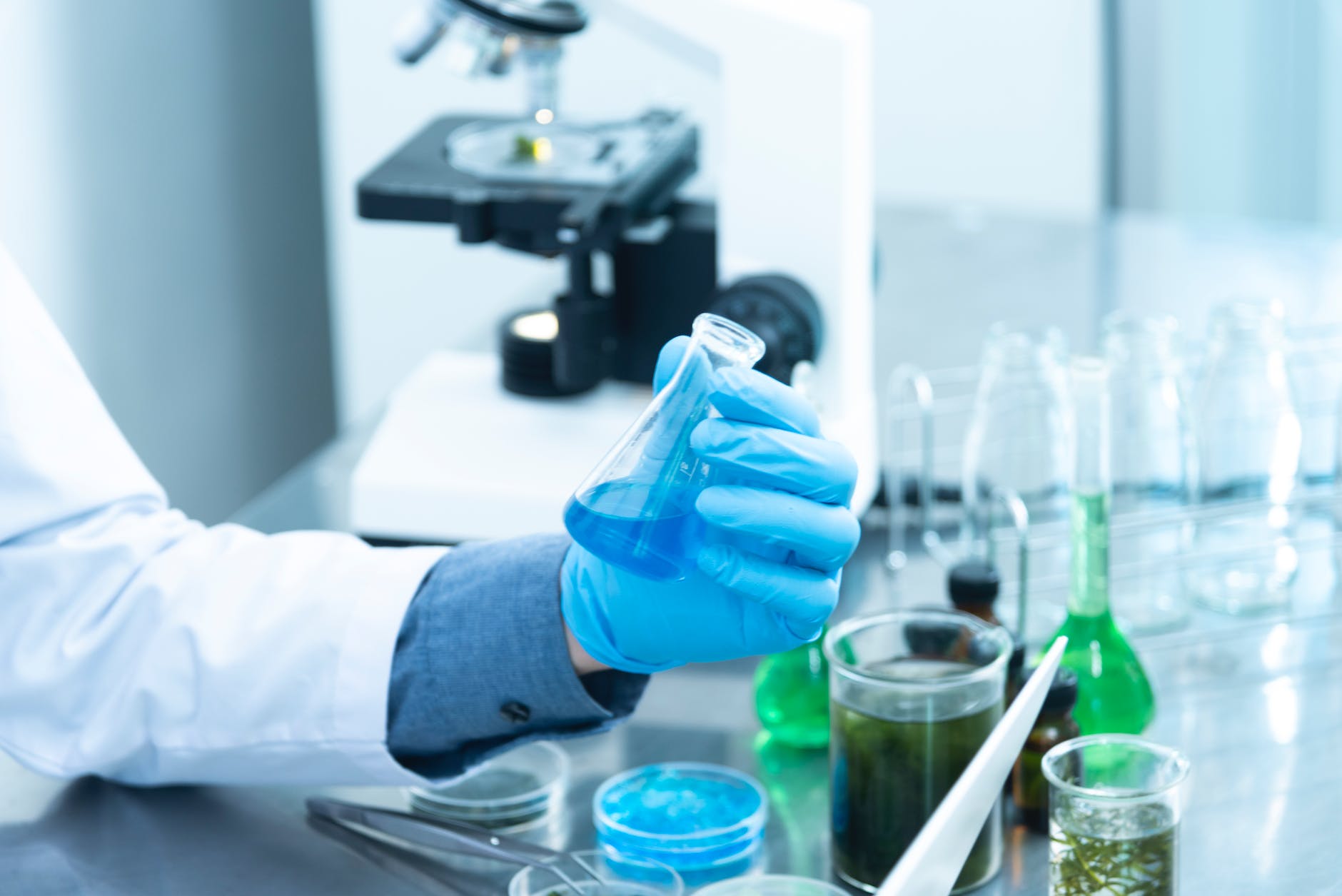
Laminar flow hood, Incubator, Autoclave, Refrigerator, Bunsen Burner, Wire loop, Petri plates, Glass slides, Weighing balance, Media plates, Sensitivity disks, Staining rack, Microscope, Bio safety Cabinet, Centrifuge etc.

INTRODUCTION TO DIFFERENT MEDIA
Some of the media used in the microbiology lab are :

  1. MacCONKEY AGAR : It is the selective and differential media used for the isolation of Gram-negative Bacteria. This media can be used for differentiating Lactose fermenting and Non-lactose fermenting bacteria.
  2. BLOOD AGAR : It is the enriched media for the growth of bacteria such as streptococci.
  3. CHOCOLATE AGAR : It is the lysed Blood Agar. The only difference in blood agar and chocolate agar is that in blood agar RBCs are lysed. This enriched media is suitable for the growth of bacteria that are unable to grow on Blood Agar.
  4. THIOSULFATE CITRATE BILE SALT AGAR (TCBS) : It is the selective as well as differential media for the growth of vibrio cholerae , a causative organism for cholera.

GRAM STAINING
Gram staining is the process for differentiating Gram positive and Gram negative bacteria. When the whole procedure of gram stain is followed and the slide is observed under the microscope, Gram positive bacteria appear Violet in color and Gram negative bacteria appear Pink in color.
For the gram staining we need Glass slide, Normal Saline, Inoculating loop, Bunsen burner, Crystal Violet, Gram’s Iodine, Acetone, Safranine.

PROCESS :

  1. The isolated colony of the microorganism is taken and in the drop of normal saline on the glass slide the colony is mixed with the help of inoculating loop to make a smear. The prepared smear is heat fixed.
  2. A staining rack is taken and on the smear, Crystal Violet is added. After 1 minute, the stain was removed by washing the slide in running water.
  3. After that, Gram’s Iodine is added on the smear as a decolorizing agent which is again washed after 1 minute under the running tap water.
  4. The next step is to add Acetone on the smear which is added in the hand to hand process.
  5. After the decolorisation is done, Safranine is added on the smear which is also washed after 1 minute.
  6. The glass slide is then air dried and observed under the microscope.

RESULTS :
It was observed under the microscope that the Gram positive bacteria appear Violet in color due to Crystal Violet stain whereas Gram negative bacteria appear Pink in color due to safranine.

TESTS ANALYZED
The tests analyzed in the microbiology section of the laboratory are basically the culture and sensitivities tests of urine, stool, sputum, pus swab etc.
The basic procedure of performing all the tests are :

  1. First of all, all the tests are performed inside the laminar flow hood.
  2. The samples collected from the patients and the media plates are kept inside the laminar flow.
  3. The inoculating wire loop is heat sterilized and with the help of it, the samples are cultured or streaked on the media plates.
  4. After inoculation, the cultured media plates are incubated for 24 hours (48 hours if necessary) for allowing the growth of bacteria.
  5. After the growth, staining is done or sensitivities are checked according to the requirement by the doctor.
  6. The report is prepared for the patient.