Discovery and working of an MRI

      The MRI (Magnetic resonance imaging) scan is a medical imaging procedure that uses a magnetic field and radio waves to take pictures of our body’s interior. It is mainly used to investigate or diagnose the conditions that affect soft tissue such as tumors or brain disorders. The MRI scanner is a complicated piece of equipment that is expensive to use and found only in specialized centers. Although Raymond Vahan Damadian (1936) is credited with the idea of turning nuclear magnetic resonance to look inside the human body, it was Paul Lauterbur (1929-2007) and Peter Mansfield (1933) who carried out the work most strongly linked to Magnetic resonance imaging (MRI) technology. The technique makes use of hydrogen atoms resonating when bombarded with magnetic energy. MRI provides three dimensional images without harmful radiation and offers more detail than older techniques.

       While training as a doctor in New York, Damadian started investigating living cells with a nuclear magnetic resonance machine. In 1971 he found that the signals carried on for longer with cells from tumors than from healthy ones. But the methods used at this time were neither effective nor practical although Damadian received a patent for such a machine to be used by doctors to pick up cancer cells in 1974.

The first full body MRI scanner at the University of Aberdeen in Scotland (1970)

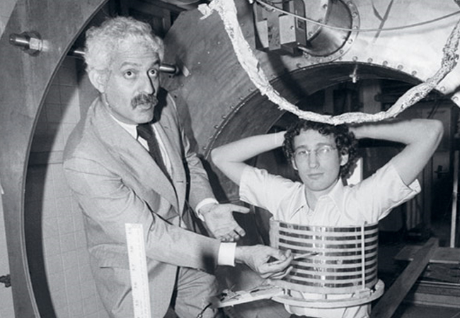
       The real shift came when Lauterbur, a U.S, chemist, introduced gradients to the magnetic field so that the origin of radio waves from the nuclei of the scanned object could be worked out. Through this he created the first MRI images in two and here dimensions. Mansfield, a physicist from England, came up with a mathematical technique that would speed up scanning and make clearer images. Damadian went on to build the full body MRI machine in 1977 and he produced the first full MRI scan of the heart, lungs, and chest wall of his skinny graduate student, Larry Minkoff – although in a very different way to modern imaging.

Working of an MRI machine

        The key components of an MRI machine are magnet, radio waves, gradient, and a super advanced computer. We all know that human bodies are made up of 60% water, and water is magnetic. Each of the billons of water molecules inside us consists of an oxygen atom bonded to two hydrogen atoms that are called as H2O. Small parts of the hydrogen atoms act as tiny magnets and are very sensitive to magnetic fields. The first step in taking an MRI scan is to use a big magnet to produce a unified magnetic field around the patient. The gradient adjusts the magnetic field into smaller sections of different magnetic strengths to isolate our body parts. Take brain as an example, normally the water molecules inside us are arranged randomly. But when we lie inside the magnetic field, most of our water molecules move at the same rhythm or frequency as the magnetic field. The ones that don’t move along the magnetic field are called low energy water molecules. To create an image of a body part, the machine focuses on the low energy molecules. The radio waves move at the same rhythm or frequency as the magnetic fields in an MRI machine.

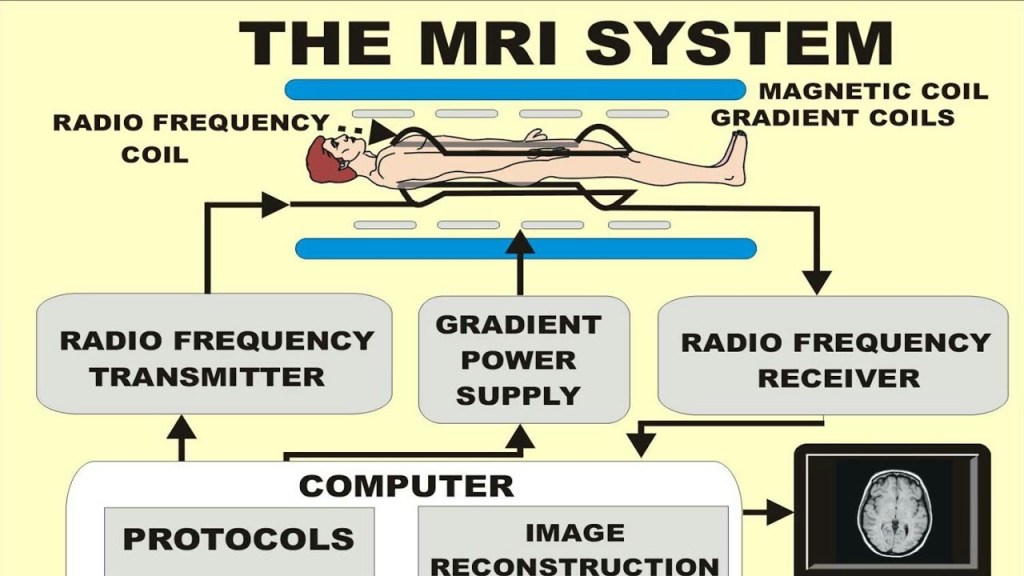
       By sending radio waves that match or resonate with the magnetic field, the low energy water molecules absorb the energy they need to move alongside the magnetic field. When the machine stops emitting radio waves, the water molecules that had just moved along the magnetic field release the energy they had absorbed and go back to their position. This movement is detected by the MRI machine and the signal is sent to a powerful computer which uses imaging software to translate the information into an image of the body. By taking images of the body in each section of the magnetic field the machine produces a final three dimensional image of the organ which doctors can analyze to make a diagnosis.

“Medicine is a science of uncertainty and an art of probability”. –William Osler

Trials of a more precise and comfortable MRI to identify early breast cancer begins in the United Kingdom.

A new, more convenient method of detecting breast cancer has begun testing in the United Kingdom, with the potential to identify tumors at an earlier stage.

At some time in their life, one in every eight British women will be diagnosed with breast cancer. Because malignancies and fibrous tissue both show up as solid white regions on X-ray, mammograms are less effective in younger women whose breasts contain denser, fibrous tissue and less fat because cancers and fibrous tissue both appear as solid white areas on X-ray.

Mammograms may miss malignancies in postmenopausal women with thick breast tissue, who are also more likely to acquire breast cancer in the first place.

Women with a dubious lump may be given an ultrasound scan or a biopsy, and if the diagnosis is still unclear, they may be directed for DCE-MRI, which detects the development of new blood vessels supporting tumors. However, in women with early-stage malignancies, they may not be visible, leading to false comfort.

Multiparametric MRI was first created to evaluate liver disorders without the need for a painful biopsy, and it is now widely utilized in Europe and the United States.

It works similarly to traditional MRI in that it utilizes high magnetic fields and radio waves to excite protons in the tissue, then exploits variations in the time it takes for them to settle to generate a “map” of the various tissues in the breast. Multiparametric MRI, on the other hand, allows an even more comprehensive map to be generated by merging pictures created by multiple MR pulses and sequences.

A scan of healthy breasts with multiparametric MRI. Photograph courtesy of Perspectum Diagnostics

“We believe that if we differentiate the tissue rather than looking at the blood vessels around the tumor, we should be able to spot not only tumors in dense breasts but potentially tumors that aren’t seen on mammograms,” said Prof Sally Collins, a consultant obstetrician and medical lead for women’s health at Oxford-based Perspectum Diagnostics, who herself recently received treatment for breast cancer.

“We’re also working to improve the scanning experience for patients. “Mammograms are awful because they squish your breast on this plate, which is undignified and unpleasant, and MRIs are much worse because you have to lie face down with your boobs dangling in this coil and your arms raised over your head for ages,” Collins added.

“We’re attempting to make it such that women may be fully dressed, respectable, and comfortable while being scanned, which is critical for the patient journey to cancer diagnosis.”

The prospectus has received ethical clearance to enroll 1,030 women in the study, including 10 women who have been diagnosed with breast cancer and 30 to 40 healthy women who are presently being scanned, to see if the technology can properly map their breast tissue while laying on their backs. It is estimated that the study would take two years to complete.

“It will never replace regular mammography screening for postmenopausal women, but we think it will enhance the diagnosis route for women with thick breasts or premenopausal women who are at extremely high risk of breast cancer, eliminating the need for repeated tests,” Collins said.

MRI

As medical sciences is getting better everyday it is mostly focussing more and more on the specific organ or part of the body having problem rather than doing a trial and error medication. For this it is really important that the doctors have the knowledge of the specific area of problem and body scanners do serves this purpose. They scan the body and develops the image of the inner body organs. There are many scanners out there like

  1. X- rays – This is the most common method used to scan the injured bones, the rays are used as they are blocked by bones and dense tissues of the body.
  2. CT scan – Also called computed tomography this is used to get detailed view of blood vessels and  organs in the body.

Third most important scanning technique is the MRI whose full form is  magnetic resonance imaging, the technique was invented in 1972 by Felix Bloch and Edward Purcell. Although in the early years this scanning technique was not used in human body, perhaps this idea was put by Raymond Damadian as he showed the doctors a way to actually use MRI to detect tumors. After some years of developing the machine so as to use it on human, in 1977 it was the first time that MRI was used on humans.

Physics behind MRI scanning

MRI is claimed to be better than CT scanning as it gives more detailed images of the body parts. Another important difference is MRI do not use radiations as CT or X-rays, as exposure to radiations can be a bit risky and especially X-rays can be harmful for pregnant women.

As we know about 60% percent of our body is  composed of water and chemical composition of water contains 2 hydrogen atoms and one oxygen atom. The hydrogen atom has one proton and one electron, and the nuclei has a characteristic  precession  because of which the magnetic moment  is created. when the body is exposed to a magnetic field of 1.5 to 3 Tesla, these magnetic moment aligns to the direction of magnetic field as the nuclei starts precessing about the magnetic field they are exposed to, this precession is called larmor precession. The frequency of the precession is proportional to the applied magnetic field. Next a radio frequency pulse is applied  which is equal to the frequency of the larmor precession, this causes the energy exchange and as they are in resonance. This causes the direction of magnetic moment to tilt a bit in the direction of the radio frequency pulse applied and when this radio frequency pulse is removed it tends magnetic moment to again come back to its position that is to align in the direction of the magnetic field . As this happens the nuclei emits its own radio frequency pulse, and different tissues in the body gives out different Radio frequency pulse. To detect this pulse  the body is covered by a coil which detects these pulses and these are converted to an image using the computers which uses mathematical methods like fourier transformation. This results in the highly accurate image of the body parts.

Though MRI is simple and has less risks like all these years pregnant women take MRI scans and there has not been any report of  any abnormalities while the child is born, but there are some precautions to be taken as follows

  1. Make sure one does not have any magnetic objects while going into the room
  2. There are many patients who fall sick after MRI, it is really normal because of the sedative given before the scan
  3. One should never move while MRI scan is been taken.
  4.  One should avoid driving after he or she has taken  MRI scan.